4. Una forma aferrada
4. A Clinging Form
Luego hicimos de aquella gota de fluido una forma adherida.
Then We made that drop of fluid into a clinging form
El Corán describe la siguiente etapa del embrión humano en desarrollo con la palabra ʿalaqah. Esta palabra tiene diversos significados, entre ellos: colgar, estar suspendido, ser colgado, pegarse, aferrarse, adherirse y unirse. También puede significar atrapar, ser atrapado, estar fijado o anexado.48
The Qur’an describes the next stage of the developing human embryo with the word `alaqah. This word carries various meanings including: to hang, to be suspended, to be dangled, to stick, to cling, to cleave and to adhere. It can also mean to catch, to get caught, to be affixed or subjoined.48
Otras connotaciones de la palabra 'alaqah incluyen una sustancia parecida a la sanguijuela, con la semejanza de un gusano; o tener una disposición 'rastrera', inclinada a succionar sangre. Por último, su significado incluye el barro que se adhiere a la mano y la sangre espesa y coagulada, por su tendencia a aferrarse entre sí. 49
Other connotations of the word `alaqah include a leech-like substance, having the resemblance of a worm; or being of a ‘creeping’ disposition inclined to the sucking of blood. Finally, its meaning includes clay that clings to the hand and thick, clotted blood - because of its clinging together. 49
INTERPRETACIÓN CIENTÍFICA
SCIENTIFIC INTERPRETATION
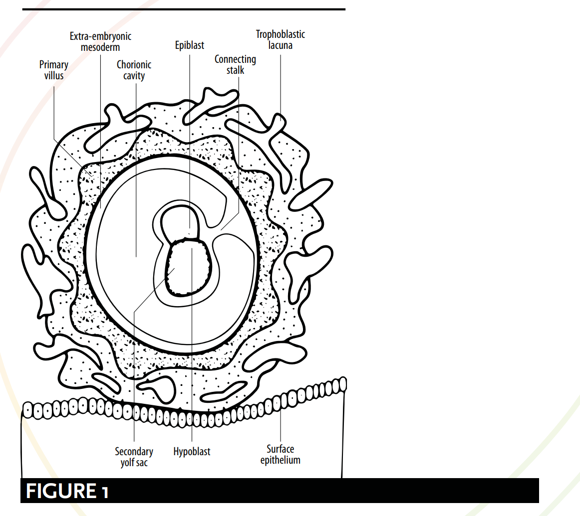
Según la embriología moderna, los múltiples significados de la palabra *'alaqah* corresponden a las distintas etapas del desarrollo del embrión. Pueden establecerse comparaciones entre las descripciones coránicas y científicas de la apariencia del embrión y de su relación con el útero. De acuerdo con la embriología moderna, a partir del día 15 el embrión queda colgado o suspendido mediante el «tallo de conexión» [véase la Figura 1] y obtiene nutrientes por contacto con los vasos sanguíneos maternos. Esta descripción guarda un sorprendente parecido con la imagen que transmite la palabra *'alaqah*: una sustancia «colgante» o «suspendida» que obtiene su alimento de la sangre de su huésped.
As defined by modern embryology, the myriad of meanings for the word `alaqah correspond to various stages of the embryo’s development. Comparisons can be drawn between qu’ranic and scientific depictions of the embryo's appearance and its relationship with the womb. According to modern embryology, from day 15 the embryo is hanging or suspended via the ‘connecting stalk’ [see Figure 1] and it obtains nutrients through contact with the maternal blood vessels. This description bears a striking resemblance to the picture painted by the word `alaqah – a ‘hanging’ or ‘suspended’ substance, obtaining nutrients from its host’s blood.
Tras esto, durante la cuarta semana, se producen dos procesos: la formación del cerebro y de la médula espinal, conocida como neurulación; y las etapas iniciales del plegamiento del embrión. Es al culminar estos dos procesos cuando el embrión adquiere una forma semejante a una sanguijuela, la forma sugerida por la palabra `alaqah: una sustancia reptante, parecida a una sanguijuela o a un gusano [véanse las Figuras 2, 3 y 4].
Following this, during the 4th week, two processes occur: the formation of the brain and the spinal cord, known as neurulation; and the initial stages of the folding of the embryo. It is upon the culmination of these two processes that the embryo resembles a leech-like form suggested by the word `alaqah – a creeping, leech or worm-like substance [see Figure 2, 3 and 4].
AHORCAMIENTO/SUSPENDIDO
HANGING/SUSPENDED
Los embriólogos Barry Mitchell y Ram Sharma explican los aspectos "colgante" o "suspendido" de la etapa de `alaqah. Describen al embrión como "conectado al citotrofoblasto por un tallo conector de mesodermo extraembrionario (tejido conectivo primitivo). El tallo es el precursor del cordón umbilical". 50 La Figura 1 muestra el embrión conectado al citotrofoblasto mediante un tallo conector, como si estuviera colgando o suspendido.
Embryologists Barry Mitchell and Ram Sharma explain the ‘hanging’ or ‘suspended’ aspects of the `alaqah stage. They describe the embryo as being, “connected to the cytotrophoblast by a connecting stalk of extra-embryonic mesoderm (primitive connective tissue). The stalk is the forerunner of the umbilical cord.” 50 Figure 1 shows the embryo connected to the cytotrophoblast via a connecting stalk, as if it were hanging or suspended.
OBTENCIÓN DE NUTRIENTES A TRAVÉS DE LOS VASOS SANGUÍNEOS
OBTAINING NUTRIENTS VIA BLOOD VESSELS
Con respecto a la forma en que el embrión obtiene sus nutrientes mediante el contacto con los vasos sanguíneos maternos, Barry Mitchell y Ram Sharma escriben: Debido al rápido crecimiento del embrión durante la segunda semana, se necesita un medio más eficiente para el intercambio de nutrientes y gases. Esto se logra cuando los vasos sanguíneos embrionarios del corion entran en contacto con los vasos sanguíneos maternos de la decidua.51.
With regard to the way the embryo obtains its nutrients through contact with the maternal blood vessels, Barry Mitchell and Ram Sharma write: Due to the rapid growth of the embryo during the second week, there is a need for a more efficient means of nutritional and gaseous exchange. This is achieved when the embryonic blood vessels of the chorion come into contact with the maternal blood vessels of the decidua.51.
... [para la tercera semana]... El intercambio de nutrientes, gases respiratorios y productos de desecho entre la sangre materna y la sangre fetal se lleva a cabo a través de la membrana placentaria, dentro de los espacios intervellosos. La sangre materna entra en estos espacios desde las arterias espirales, ramas de la arteria uterina, aportando nutrientes y oxígeno para el embrión y el feto.52
... [by the third week]... The exchange of nutrients, respiratory gases and waste products between the maternal and fetal blood takes place across the placental membrane within intervillous spaces. Maternal blood enters these spaces from the spiral arteries, branches of the uterine artery, bringing nutrients and oxygen for the embryo and fetus.52
Esto refuerza la validez de los significados de 'alaqah, y su evocación de imágenes de una entidad que obtiene sus nutrientes a través de la sangre.
This reinforces the validity of the meanings of `alaqah, and its evoking images of an entity obtaining its nutrients via blood.
NEURULACIÓN Y PLEGAMIENTO DEL EMBRIÓN
NEURULATION & THE FOLDING OF THE EMBRYO
La etapa de la `alaqah sugiere el proceso de neurulación y las etapas iniciales del plegamiento del embrión. La neurulación comprende la formación del cerebro y de la médula espinal entre los días 19 y 25 (aproximadamente); y el plegamiento del embrión implica que la cabeza y la cola se acerquen entre sí. La combinación de estos dos cambios fisiológicos hace que el embrión se asemeje a una sustancia parecida a una sanguijuela.
The `alaqah stage suggests the process of neurulation and the initial stages of the folding of the embryo. Neurulation comprises of the formation of the brain and the spinal cord from days 19 to 25 (approx.); and the folding of the embryo involves the head and the tail being brought closer together. The combination of these two physiological changes causes the embryo to resemble a leech-like substance.
NEURULACIÓN
NEURULATION
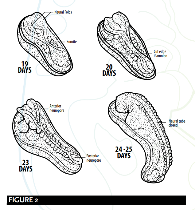
Al final de la neurulación se cierran los extremos craneal y caudal del tubo neural. En este punto, el embrión adquiere un aspecto similar al de una sanguijuela 53 [véase la Figura 2].
At the end of neurulation the cranial and caudal ends of the neural tube close. The embryo, at this point, becomes leech-like in appearance 53 [see Figure 2].
Barry Micthell y Ram Sharma explican el proceso de neurulación.
Barry Micthell and Ram Sharma explain the process of neurulation.
Hacia los 19 días, en el extremo craneal de la línea primitiva, el mesodermo subyacente y la notocorda inducen al ectodermo a formar la placa neural, que se redondea para formar los pliegues neurales. La placa neural se agranda inicialmente en el extremo craneal. A los 20 días, la placa neural en la región media del embrión permanece estrechada, pero se expande en el extremo caudal. La placa se profundiza para formar el surco neural, a partir del cual se forma el tubo neural. Los extremos craneal y caudal del tubo están abiertos y se conocen como neuroporos anterior y posterior; estos finalmente se cierran. 54
At about 19 days, at the cranial end of the primitive streak, the underlying mesoderm and notochord induce the ectoderm to form the neural plate, which rounds up to form the neural folds. The neural plate enlarges initially at the cranial end. At 20 days, the neural plate in the mid-region of the embryo remains narrowed, but it expands at the caudal end. The plate deepens to form the neural groove from which the neural tube forms. The cranial and caudal ends of the tube are open and are known as the anterior and posterior neuropores; these eventually close. 54
PLEGADO DEL EMBRIÓN
FOLDING OF THE EMBRYO
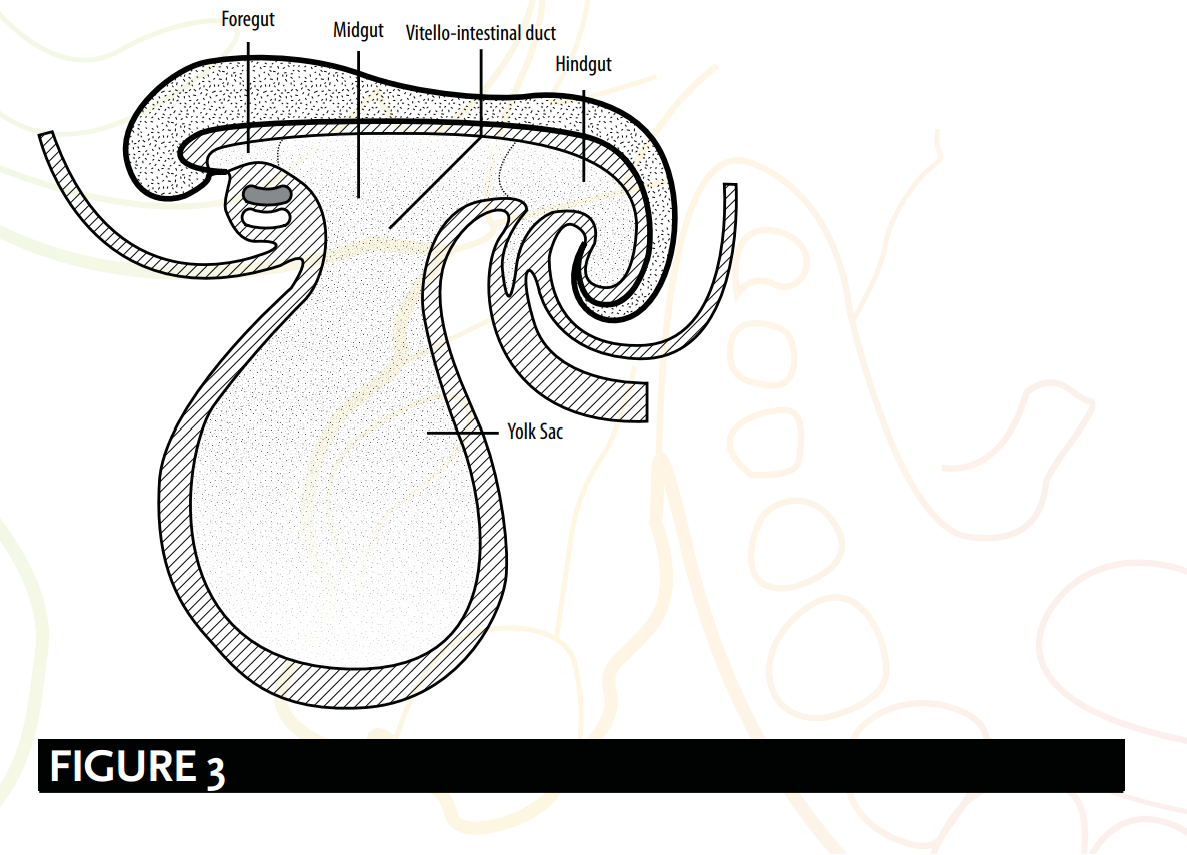
El plegamiento del embrión también es responsable de dar forma a una estructura parecida a una sanguijuela [véase la Figura 3], o, como lo han descrito los embriólogos, una estructura cilíndrica o tubular. Los embriólogos Keith Moore y T. V. N. Persaud sugieren que:
The folding of the embryo is also responsible for forming a leech-like shape [see Figure 3], or as embryologists have described; a cylindric or tube-like structure. Embryologists Keith Moore and T. V. N. Persaud suggest that:
Un acontecimiento significativo en el establecimiento de la forma corporal es el plegamiento del disco embrionario trilaminar plano en un embrión algo cilíndrico. El plegamiento resulta del rápido crecimiento del embrión, en particular del encéfalo y de la médula espinal. 55
A significant event in the establishment of body form is the folding of the flat trilaminar embryonic disc into a somewhat cylindric embryo. Folding results from the rapid growth of the embryo, particularly the brain and the spinal cord. 55
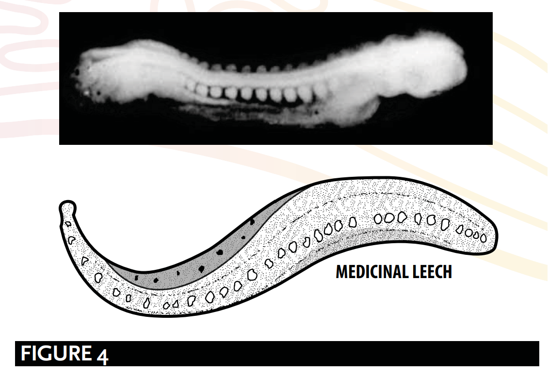
La estructura en forma de tubo o de sanguijuela [ver Figura 4], tal como la explican Barry Mitchell y Ram Sharma, se debe a:
The tube-like or leech-like structure [see Figure 4], as explained by Barry Mitchell and Ram Sharma, is due to:
El plegamiento longitudinal, que ocurre entre los días 21 y 24, resulta en… el embrión se curva de modo que la cabeza y la cola se acercan entre sí… para formar una estructura con forma de tubo. 56
longitudinal folding, which occurs between days 21 and 24, result[ing] in … the embryo [bending] so that the head and tail are brought closer together...[to] form a tube-like structure. 56
RESUMEN
SUMMARY
La etapa de la ʻalaqah:
The `alaqah stage:
El embrión está conectado al citotrofoblasto mediante un pedúnculo de unión, como si estuviera colgado o suspendido.
The embryo is connected to the cytotrophoblast via a connecting stalk, as if it were hanging or suspended.
La formación del cerebro y de la médula espinal, conocida como neurulación, y durante las etapas iniciales del plegamiento del embrión. Es al culminar estos procesos cuando el embrión adquiere apariencia de sanguijuela.
The formation of the brain and the spinal cord, known as neurulation; and during the initial stages of the folding of the embryo. It is upon the culmination of these processes that the embryo appears leech-like.
El embrión obtiene sus nutrientes a través del contacto con los vasos sanguíneos maternos. Esto refleja la acción de una sustancia similar a una sanguijuela que obtiene sus nutrientes de la sangre.
The embryo obtains its nutrients via contact with the maternal blood vessels. This mirrors the action of a leech-like substance obtaining its nutrients via blood.
GALENO Y ‘ALAQAH
GALEN & `ALAQAH
Los críticos afirman que el uso de la palabra ʿalaqah es una descripción de la segunda etapa del embrión humano según Galeno. Su traducción de la palabra ʿalaqah como "coágulo de sangre" crea la impresión de que se trata de la descripción de Galeno de esta etapa.
Critics declare that the use of the word `alaqah is a description of Galen’s second stage of the human embryo. Their translation of the word `alaqah to ‘blood clot’ creates an impression of Galen's description of this stage:
Pero cuando [el coágulo] se ha llenado de sangre, y el corazón, el cerebro y el hígado todavía no están articulados ni formados, pero ya tienen cierta solidez y un tamaño considerable, este es el segundo período; la sustancia del feto tiene la forma de carne y ya no la forma de semen. 57
But when it [the clot] has been filled with blood, and heart, brain and liver are still unarticulated and unshaped yet have by now certain solidarity and considerable size, this is the second period; the substance of the foetus has the form of flesh and no longer the form of semen. 57
Hay varias debilidades en las implicaciones de estos críticos:
There are a number of weaknesses with the implications of these critics:
Lamentablemente, el texto completo no llegó; solo recibí el inicio: "1. Firstly, the Qur'an does not make a mention of the 'heart, brain and liver...". Por favor, envíe el resto del pasaje para traducirlo.
1. Firstly, the Qur’an does not make a mention of the “heart, brain and liver...
««sin articular y sin forma todavía, pero con cierta solidaridad y de un tamaño considerable»
[being] unarticulated and unshaped yet [with] a certain solidarity and [of a] considerable size”.
2. En segundo lugar, la palabra ʿalaqah no significa sangre; más bien, sus significados principales son «una forma que se adhiere» y «una sustancia semejante a una sanguijuela». Algunos lingüistas árabes atribuyen a la palabra ʿalaqah el significado de «coágulo de sangre», no porque esté llena de sangre, sino por su naturaleza de «adherirse». Esta interpretación coincide con el significado primario de la palabra.
2. Secondly, the word `alaqah does not mean blood; rather, its primary meanings are ‘a clinging form’ and ‘a leech-like substance’. Some Arab linguists would attribute the meaning of a ‘blood clot’ to the word `alaqah, not because of it being filled with blood, but instead due to it ‘clinging’ by nature. This interpretation matches the primary meaning of the word.
3. En tercer lugar, Galeno insiste en que «el feto tiene la forma de carne» durante la segunda etapa, mientras que el Corán afirma que la etapa del mudghah (un trozo de carne masticada o un bulto de carne) viene después de la etapa del `alaqah.
3. Thirdly, Galen insists “the foetus has the form of flesh” during the second stage, whereas the Qur’an affirms the mudghah (a chewed piece of meat or lump of flesh) stage comes after the `alaqah stage.
4. En cuarto lugar, en su libro Sobre las facultades naturales, Galeno escribe: así ocurre con el semen: sus facultades las poseía desde el principio.58 La implicación incorrecta de Galeno, según la cual el semen es el único responsable del material genético, es incompatible con el Corán y las tradiciones proféticas, y se opone directamente a ellos. Como se explicó anteriormente (véase Aristóteles, Galeno y Nutfah), los textos fuente islámicos consideran el nutfah como una gota o fluido mezclado procedente tanto del varón como de la mujer, y confirman que tanto el varón como la mujer son responsables de la genética del niño.59 Por lo tanto, si el Profeta ﷺ copió las opiniones de Galeno sobre el desarrollo humano, ¿cómo es que rechazó la ciencia incorrecta y solo utilizó lo que era correcto, aunque en la Arabia del siglo VII no había forma de saber qué opiniones eran acertadas?
REPRESENTACIÓN ERRÓNEA DE LA APARIENCIA DEL EMBRIÓN
MISREPRESENTING THE EMBRYO’S APPEARANCE
Los comentaristas contemporáneos sostienen que el embrión solo se parece a una sanguijuela cuando se retira el saco vitelino, y que, por tanto, la descripción de la ʿalaqah es una representación errónea de la apariencia del embrión en esta etapa. Sin embargo, los embriólogos explican que el saco vitelino está separado del embrión por una estructura extraembrionaria, llamada celoma extraembrionario. Este hecho refuta las afirmaciones de representación errónea, ya que el saco vitelino no forma parte de la estructura del embrión.60
Contemporary commentators argue the embryo only looks leech-like when the yolk sac is removed, and that the `alaqah's description is therefore a misrepresentation of the embryo’s appearance at this stage. However, embryologists explain that the yolk sac is separated from the embryo by an extra-embryonic feature, called the extra-embryonic coelom. This fact disproves claims of misrepresentation as the yolk sac is not part of the structure of the embryo.60