4. ایک چمٹنے والی شکل
4. A Clinging Form
پھر ہم نے اس قطرے کو جونک کی طرح چپکنے والی شکل میں بنا دیا۔
Then We made that drop of fluid into a clinging form
قرآن کریم انسانی جنین کے ارتقائی مرحلے کی اگلی حالت کو لفظ 'علقہ' سے بیان کرتا ہے۔ اس لفظ کے مختلف معانی ہیں جن میں شامل ہیں: لٹکنا، معلق ہونا، جھولنا، چپکنا، چمٹنا، جڑنا اور جڑ جانا۔ اس کے دیگر معانی میں پکڑنا، پکڑا جانا، منسلک ہونا یا جڑنا بھی شامل ہو سکتا ہے۔48
The Qur’an describes the next stage of the developing human embryo with the word `alaqah. This word carries various meanings including: to hang, to be suspended, to be dangled, to stick, to cling, to cleave and to adhere. It can also mean to catch, to get caught, to be affixed or subjoined.48
لفظ 'علقہ' کے دیگر معانی میں ایسی چیز شامل ہے جو جونک کی طرح ہو اور کیڑے سے مشابہت رکھتی ہو؛ یا ایسی چیز جس میں خون چوسنے کی 'رینگنے' والی طبیعت پائی جاتی ہو۔ آخر میں، اس کے معنی میں وہ مٹی بھی شامل ہے جو ہاتھ سے چپک جائے اور گاڑھا، جما ہوا خون بھی ہے - کیونکہ وہ آپس میں جڑا ہوا ہوتا ہے۔ 49
Other connotations of the word `alaqah include a leech-like substance, having the resemblance of a worm; or being of a ‘creeping’ disposition inclined to the sucking of blood. Finally, its meaning includes clay that clings to the hand and thick, clotted blood - because of its clinging together. 49
سائنسی تشریح
SCIENTIFIC INTERPRETATION
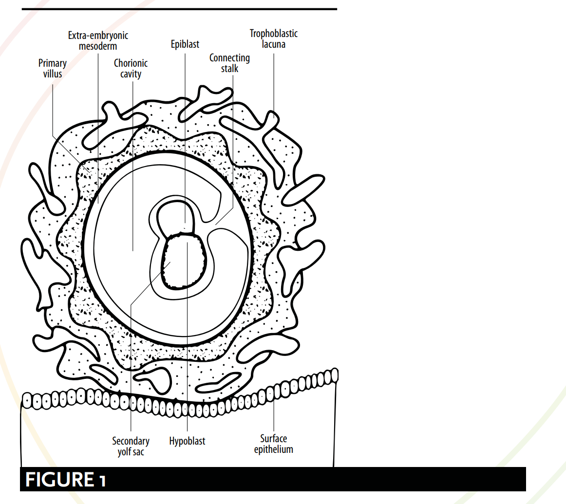
جدید جنینวิทยา (embryology) کی تعریف کے مطابق، لفظ 'علقہ' کے بے شمار معانی جنین کی نشوونما کے مختلف مراحل سے مطابقت رکھتے ہیں۔ جنین کی ظاہری شکل اور رحم کے ساتھ اس کے تعلق کے حوالے سے قرآنی اور سائنسی بیانات کے درمیان موازنہ کیا جا سکتا ہے۔ جدید جنینวิทยา کے مطابق، 15 ویں دن سے جنین 'کنیکٹنگ اسٹاک' (connecting stalk) کے ذریعے لٹکا ہوا یا معلق ہوتا ہے [دیکھیے شکل 1] اور وہ ماں کی خون کی رگوں کے ساتھ رابطے کے ذریعے غذائی اجزاء حاصل کرتا ہے۔ یہ وضاحت لفظ 'علقہ' سے پیش کردہ تصویر سے حیرت انگیز مماثلت رکھتی ہے—یعنی ایک ایسی 'لٹکی ہوئی' یا 'معلق' چیز، جو اپنے میزبان کے خون سے غذائی اجزاء حاصل کرتی ہے۔
As defined by modern embryology, the myriad of meanings for the word `alaqah correspond to various stages of the embryo’s development. Comparisons can be drawn between qu’ranic and scientific depictions of the embryo's appearance and its relationship with the womb. According to modern embryology, from day 15 the embryo is hanging or suspended via the ‘connecting stalk’ [see Figure 1] and it obtains nutrients through contact with the maternal blood vessels. This description bears a striking resemblance to the picture painted by the word `alaqah – a ‘hanging’ or ‘suspended’ substance, obtaining nutrients from its host’s blood.
اس کے بعد، چوتھے ہفتے کے دوران دو عمل رونما ہوتے ہیں: دماغ اور ریڑھ کی ہڈی کی تشکیل، جسے نیورولیشن (neurulation) کہا جاتا ہے؛ اور جنین کے تہہ ہونے (folding) کے ابتدائی مراحل۔ ان دو عملوں کے مکمل ہونے پر ہی جنین کی شکل ایک جونک جیسی ہو جاتی ہے، جیسا کہ لفظ 'علقہ' سے ظاہر ہوتا ہے— یعنی رینگنے والی، جونک یا کیڑے جیسی چیز [دیکھیے شکل 2، 3 اور 4]۔
Following this, during the 4th week, two processes occur: the formation of the brain and the spinal cord, known as neurulation; and the initial stages of the folding of the embryo. It is upon the culmination of these two processes that the embryo resembles a leech-like form suggested by the word `alaqah – a creeping, leech or worm-like substance [see Figure 2, 3 and 4].
لٹکا ہوا / معلق
HANGING/SUSPENDED
جنین کے ماہرین بیری مچل اور رام شرما 'علقہ' کے مرحلے کے 'لٹکنے' یا 'معلق' ہونے کے پہلوؤں کی وضاحت کرتے ہیں۔ وہ جنین کی وضاحت یوں کرتے ہیں کہ، "یہ ایک اضافی جنینی میسودرم (ابتدائی قمشی بافت) کے رابطے والے ڈنڈے (connecting stalk) کے ذریعے سائٹو ٹروفوبلاسٹ سے جڑا ہوتا ہے۔ یہ ڈنڈا ناف کی رسی کا پیش رو ہے۔" 50 شکل 1 میں جنین کو ایک رابطے والے ڈنڈے کے ذریعے سائٹو ٹروفوبلاسٹ سے جڑا ہوا دکھایا گیا ہے، گویا کہ وہ لٹکا ہوا یا معلق ہو۔
Embryologists Barry Mitchell and Ram Sharma explain the ‘hanging’ or ‘suspended’ aspects of the `alaqah stage. They describe the embryo as being, “connected to the cytotrophoblast by a connecting stalk of extra-embryonic mesoderm (primitive connective tissue). The stalk is the forerunner of the umbilical cord.” 50 Figure 1 shows the embryo connected to the cytotrophoblast via a connecting stalk, as if it were hanging or suspended.
خون کی رگوں کے ذریعے غذائی اجزاء کا حصول
OBTAINING NUTRIENTS VIA BLOOD VESSELS
اس حوالے سے کہ بھروا (embryo) ماں کی خون کی رگوں کے ساتھ رابطے کے ذریعے اپنے غذائی اجزاء کیسے حاصل کرتا ہے، بیری مچل اور رام شرما لکھتے ہیں: دوسرے ہفتے کے دوران بھروا کی تیز رفتار نشوونما کی وجہ سے، غذائی اور گیسی تبادلے کے لیے ایک زیادہ مؤثر ذریعے کی ضرورت ہوتی ہے۔ یہ اس وقت حاصل ہوتا ہے جب کورین (chorion) کی بھروا کی خون کی رگیں ڈیسڈوا (decidua) کی ماں کی خون کی رگوں کے ساتھ رابطے میں آتی ہیں۔51
With regard to the way the embryo obtains its nutrients through contact with the maternal blood vessels, Barry Mitchell and Ram Sharma write: Due to the rapid growth of the embryo during the second week, there is a need for a more efficient means of nutritional and gaseous exchange. This is achieved when the embryonic blood vessels of the chorion come into contact with the maternal blood vessels of the decidua.51.
... [تیسرے ہفتے تک]... ماں کے خون اور جنین کے خون کے درمیان غذائی اجزاء، سانس لینے والی گیسوں اور فضلے کے اخراج کا تبادلہ، انٹرو ویلوز سپیسز (intervillous spaces) کے اندر نیزک پلائی (placental membrane) کے ذریعے ہوتا ہے۔ ماں کا خون رحم کی شریان (uterine artery) کی شاخوں، یعنی اسپائرل آرٹریز (spiral arteries) کے ذریعے ان جگہوں پر داخل ہوتا ہے، جو جنین اور بچے کے لیے غذائی اجزاء اور آکسیجن لاتا ہے۔52
... [by the third week]... The exchange of nutrients, respiratory gases and waste products between the maternal and fetal blood takes place across the placental membrane within intervillous spaces. Maternal blood enters these spaces from the spiral arteries, branches of the uterine artery, bringing nutrients and oxygen for the embryo and fetus.52
یہ 'علقہ' کے معانی کی صحت کی تائید کرتا ہے، اور اس سے ایک ایسی ہستی کا تصور ابھرتا ہے جو خون کے ذریعے اپنی غذائیت حاصل کرتی ہے۔
This reinforces the validity of the meanings of `alaqah, and its evoking images of an entity obtaining its nutrients via blood.
نیورولیشن اور جنین کا مڑنا
NEURULATION & THE FOLDING OF THE EMBRYO
علقہ کا مرحلہ نیورولیشن (اعصابی نالی کی تشکیل) کے عمل اور جنین کے تہہ ہونے کے ابتدائی مراحل کی نشاندہی کرتا ہے۔ نیورولیشن میں تقریباً 19 سے 25 دنوں کے دوران دماغ اور ریڑھ کی ہڈی کی تشکیل شامل ہے؛ اور جنین کے تہہ ہونے کے عمل میں سر اور دم کا ایک دوسرے کے قریب آنا شامل ہے۔ ان دو جسمانی تبدیلیوں کے ملاپ سے جنین کی شکل ایک جونک جیسی چیز سے مشابہت اختیار کر لیتی ہے۔
The `alaqah stage suggests the process of neurulation and the initial stages of the folding of the embryo. Neurulation comprises of the formation of the brain and the spinal cord from days 19 to 25 (approx.); and the folding of the embryo involves the head and the tail being brought closer together. The combination of these two physiological changes causes the embryo to resemble a leech-like substance.
نیورولیشن (اعصابی نالی کی تشکیل)
NEURULATION
نیورولیشن (neurulation) کے اختتام پر، اعصابی نالی (neural tube) کے سریری اور دم دار سرے بند ہو جاتے ہیں۔ اس مرحلے پر، جنین کی شکل لیچ (leech) جیسی ہو جاتی ہے 53 [شکل 2 دیکھیں]۔
At the end of neurulation the cranial and caudal ends of the neural tube close. The embryo, at this point, becomes leech-like in appearance 53 [see Figure 2].
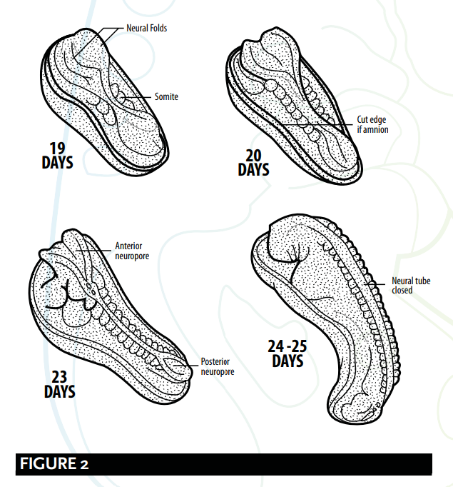
بیری مِچل اور رام شرما نیورولیشن (neurulation) کے عمل کی وضاحت کرتے ہیں۔
Barry Micthell and Ram Sharma explain the process of neurulation.
تقریباً 19 دن پر، پرائمٹیو اسٹریک (primitive streak) کے کرینئیل (cranial) سرے پر، نیچے موجود میسودرم (mesoderm) اور نوٹوکارڈ (notochord) ایکٹوڈرم (ectoderm) کو نیورل پلیٹ (neural plate) بنانے کے لیے مائل کرتے ہیں، جو کہ نیورل فولڈز (neural folds) بنانے کے لیے گول شکل اختیار کر لیتی ہے۔ نیورل پلیٹ ابتدا میں کرینئیل سرے پر پھیلتی ہے۔ 20 دن پر، جنین کے درمیانی حصے میں نیورل پلیٹ تنگ رہتی ہے، لیکن یہ کیوڈل (caudal) سرے پر پھیل جاتی ہے۔ یہ پلیٹ گہری ہو کر نیورل گروو (neural groove) بناتی ہے جس سے نیورل ٹیوب (neural tube) تشکیل پاتی ہے۔ ٹیوب کے کرینئیل اور کیوڈل سرے کھلے ہوتے ہیں جنہیں اینٹیریئر (anterior) اور پوسٹیریئر (posterior) نیوروپورز (neuropores) کہا جاتا ہے؛ یہ بالآخر بند ہو جاتے ہیں۔ 54
At about 19 days, at the cranial end of the primitive streak, the underlying mesoderm and notochord induce the ectoderm to form the neural plate, which rounds up to form the neural folds. The neural plate enlarges initially at the cranial end. At 20 days, the neural plate in the mid-region of the embryo remains narrowed, but it expands at the caudal end. The plate deepens to form the neural groove from which the neural tube forms. The cranial and caudal ends of the tube are open and are known as the anterior and posterior neuropores; these eventually close. 54
جنین کا مڑنا (Folding)
FOLDING OF THE EMBRYO
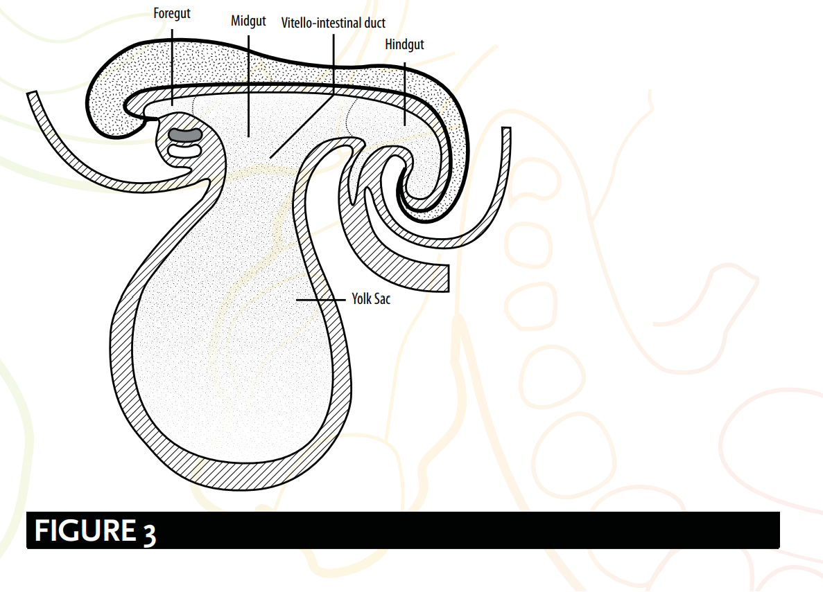
جنین کا مڑنا (folding) لیچ (leech) جیسی شکل بنانے کا بھی ذمہ دار ہے [دیکھیے شکل 3]، یا جیسا کہ جنین ماہرین نے بیان کیا ہے؛ ایک سلنڈر نما یا ٹیوب نما ساخت۔ جنین ماہرین کیتھ مور اور ٹی وی این پرسود تجویز کرتے ہیں کہ:
The folding of the embryo is also responsible for forming a leech-like shape [see Figure 3], or as embryologists have described; a cylindric or tube-like structure. Embryologists Keith Moore and T. V. N. Persaud suggest that:
جسمانی ساخت کے قیام میں ایک اہم واقعہ چپٹے سہ لایتی جنینی ڈسک (trilaminar embryonic disc) کا ایک حد تک سلنڈر نما جنین میں مڑ جانا ہے۔ یہ مڑنا جنین کی تیز رفتار نشوونما، بالخصوص دماغ اور ریڑھ کی ہڈی کے نتیجے میں ہوتا ہے۔ 55
A significant event in the establishment of body form is the folding of the flat trilaminar embryonic disc into a somewhat cylindric embryo. Folding results from the rapid growth of the embryo, particularly the brain and the spinal cord. 55
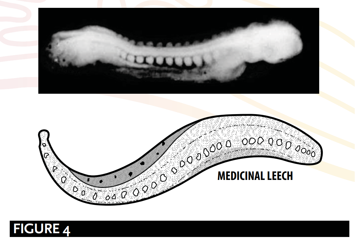
بیری مچل اور رام شرما کی وضاحت کے مطابق، نالی نما یا جونک نما ساخت [دیکھیے شکل 4] کی وجہ یہ ہے:
The tube-like or leech-like structure [see Figure 4], as explained by Barry Mitchell and Ram Sharma, is due to:
طولیہ تہوں کا عمل، جو 21 سے 24 دنوں کے درمیان ہوتا ہے، اس کے نتیجے میں... جنین اس طرح مڑ جاتا ہے کہ سر اور دم ایک دوسرے کے قریب آ جاتے ہیں... تاکہ ایک نالی نما ساخت بن سکے۔ 56
longitudinal folding, which occurs between days 21 and 24, result[ing] in … the embryo [bending] so that the head and tail are brought closer together...[to] form a tube-like structure. 56
خلاصہ
SUMMARY
علقہ کا مرحلہ:
The `alaqah stage:
جنین ایک کنیکٹنگ اسٹاک (connecting stalk) کے ذریعے سائٹوٹروفوبلاسٹ (cytotrophoblast) سے جڑا ہوا ہوتا ہے، گویا کہ وہ لٹکا ہوا یا معلق ہو۔
The embryo is connected to the cytotrophoblast via a connecting stalk, as if it were hanging or suspended.
دماغ اور ریڑھ کی ہڈی کی تشکیل، جسے نیورولیشن (neurulation) کہا جاتا ہے؛ اور یہ عمل جنین کے تہہ ہونے کے ابتدائی مراحل کے دوران ہوتا ہے۔ ان عمل کے مکمل ہونے پر ہی جنین لیچ (leech) کی مانند نظر آتا ہے۔
The formation of the brain and the spinal cord, known as neurulation; and during the initial stages of the folding of the embryo. It is upon the culmination of these processes that the embryo appears leech-like.
جنین ماں کی خون کی رگوں کے ساتھ رابطے کے ذریعے اپنی غذائی اجزاء حاصل کرتا ہے۔ یہ عمل ایک ایسی جونک نما چیز کی طرح ہے جو خون کے ذریعے اپنی غذائی اجزاء حاصل کرتی ہے۔
The embryo obtains its nutrients via contact with the maternal blood vessels. This mirrors the action of a leech-like substance obtaining its nutrients via blood.
جالین اور علاقتہ
GALEN & `ALAQAH
نقاد کا دعویٰ ہے کہ لفظ 'علقہ' کا استعمال انسانی جنین کے جالینوس کے دوسرے مرحلے کی وضاحت ہے۔ لفظ 'علقہ' کا ان کا ترجمہ 'خون کا لوتھڑا' جالینوس کی اس مرحلے کی وضاحت کا تاثر پیدا کرتا ہے:
Critics declare that the use of the word `alaqah is a description of Galen’s second stage of the human embryo. Their translation of the word `alaqah to ‘blood clot’ creates an impression of Galen's description of this stage:
لیکن جب وہ [علقہ] خون سے بھر جاتا ہے، اور دل، دماغ اور جگر ابھی تک واضح اور شکل اختیار نہیں کر پائے ہوتے مگر اب ان میں ایک خاص ٹھوس پن اور کافی حجم آ چکا ہوتا ہے، تو یہ دوسرا مرحلہ ہے؛ اس وقت جنین کا مادہ گوشت کی شکل اختیار کر لیتا ہے اور اب وہ منی کی شکل میں نہیں رہتا۔ 57
But when it [the clot] has been filled with blood, and heart, brain and liver are still unarticulated and unshaped yet have by now certain solidarity and considerable size, this is the second period; the substance of the foetus has the form of flesh and no longer the form of semen. 57
ان ناقدین کے استدلال کے نتائج میں متعدد کمزوریاں ہیں:
There are a number of weaknesses with the implications of these critics:
۱۔ اولاً، قرآن میں "دل، دماغ اور جگر..." کا ذکر نہیں ملتا...
1. Firstly, the Qur’an does not make a mention of the “heart, brain and liver...
"غیر واضح اور بے شکل، مگر ایک خاص یکجہتی اور کافی بڑے حجم کے ساتھ"۔
[being] unarticulated and unshaped yet [with] a certain solidarity and [of a] considerable size”.
۲۔ دوم ، لفظ 'علقہ' کا مطلب خون نہیں ہے؛ بلکہ اس کے بنیادی معانی 'چپکنے والی چیز' اور 'جونک جیسی چیز' ہیں۔ بعض عرب ماہرینِ لغت لفظ 'علقہ' کا مطلب 'خون کا لوتھڑا' اس لیے قرار دیتے ہیں کیونکہ وہ خون سے بھرا ہوتا ہے، بلکہ اس لیے کہ وہ اپنی فطرت کے اعتبار سے 'چپکنے' والا ہوتا ہے۔ یہ تشریح لفظ کے بنیادی معنی کے عین مطابق ہے۔
2. Secondly, the word `alaqah does not mean blood; rather, its primary meanings are ‘a clinging form’ and ‘a leech-like substance’. Some Arab linguists would attribute the meaning of a ‘blood clot’ to the word `alaqah, not because of it being filled with blood, but instead due to it ‘clinging’ by nature. This interpretation matches the primary meaning of the word.
3۔ تیسری بات یہ کہ جالینوس کا اصرار ہے کہ دوسرے مرحلے کے دوران "جنین گوشت کی شکل اختیار کر لیتا ہے"، جبکہ قرآن کریم اس بات کی تصدیق کرتا ہے کہ مضغہ (چبایا ہوا گوشت کا ٹکڑا یا گوشت کا لوتھڑا) کا مرحلہ، علقہ کے مرحلے کے بعد آتا ہے۔
3. Thirdly, Galen insists “the foetus has the form of flesh” during the second stage, whereas the Qur’an affirms the mudghah (a chewed piece of meat or lump of flesh) stage comes after the `alaqah stage.
4۔ چوتھی بات یہ کہ، جالینوس نے اپنی کتاب 'On the Natural Faculties' میں لکھا ہے: منی کا معاملہ بھی ایسا ہی ہے: اس کی قوتیں شروع سے ہی اس میں موجود تھیں۔58 جالینوس کا یہ غلط مفروضہ کہ موروثی مواد کی تمام تر ذمہ داری صرف منی پر ہے، قرآن اور احادیثِ نبوی ﷺ کے منافی اور ان کے بالکل برعکس ہے۔ جیسا کہ پہلے بحث کی جا چکی ہے (دیکھیے: ارسطو، جالینوس اور نطفہ) اسلامی بنیادی متن نطفہ کو مرد اور عورت دونوں کے ملے جلے قطرے یا مائع کے طور پر تسلیم کرتا ہے، اور اس بات کی تصدیق کرتا ہے کہ بچے کے جینیات (genetics) کے لیے مرد اور عورت دونوں ذمہ دار ہیں۔59 لہٰذا، اگر نبی ﷺ نے انسانی نشوونما کے بارے میں جالینوس کے نظریات کی نقل کی ہوتی، تو یہ کیسے ممکن ہے کہ آپ ﷺ نے غلط سائنس کو مسترد کر دیا اور صرف وہی استعمال کیا جو درست تھا، باوجود اس کے کہ ساتویں صدی کے عرب میں یہ جاننے کا کوئی ذریعہ نہ تھا کہ کون سے نظریات درست ہیں؟
4. Fourthly, in his book On the Natural Faculties, Galen writes: so it is with the semen: its faculties it possessed from the beginning.58 Galen’s incorrect implication of the semen’s sole responsibility for the genetic material is incompatible, and in direct opposition, to the Qur’an and Prophetic traditions. As previously explored (see Aristotle, Galen & Nutfah) the Islamic source texts considers the nutfah as a mingled drop or fluid from both the male and the female, and confirm both the male and female as responsible for the genetics of the child.59 Therefore, if the Prophet ﷺ copied Galen’s views on human development, how is it that he rejected the incorrect science and only used what was correct, even though there was no way in 7th century Arabia of knowing which views were accurate?
جنین کی ظاہری شکل کی غلط بیانی
MISREPRESENTING THE EMBRYO’S APPEARANCE
عصر حاضر کے مفسرین اس بات پر دلیل دیتے ہیں کہ جنین صرف اس وقت جونک کی طرح نظر آتا ہے جب صفاقی (yolk sac) کو الگ کر دیا جائے، اور اس لیے 'علقہ' کی وضاحت اس مرحلے پر جنین کی ظاہری شکل کی غلط بیانی ہے۔ تاہم، ماہرینِ جنین (embryologists) وضاحت کرتے ہیں کہ صفاقی کو جنین سے ایک غیر جنینی خصوصیت کے ذریعے الگ کیا جاتا ہے جسے 'ایکسٹرا ایمبریونک سیلوم' (extra-embryonic coelom) کہا جاتا ہے۔ یہ حقیقت غلط بیانی کے دعووں کو غلط ثابت کرتی ہے کیونکہ صفاقی جنین کی ساخت کا حصہ نہیں ہوتی۔60
Contemporary commentators argue the embryo only looks leech-like when the yolk sac is removed, and that the `alaqah's description is therefore a misrepresentation of the embryo’s appearance at this stage. However, embryologists explain that the yolk sac is separated from the embryo by an extra-embryonic feature, called the extra-embryonic coelom. This fact disproves claims of misrepresentation as the yolk sac is not part of the structure of the embryo.60