4. Eine anhaftende Form
4. A Clinging Form
Dann machten Wir jenen Tropfen Flüssigkeit zu einem sich festsetzenden Gebilde.
Then We made that drop of fluid into a clinging form
Der Koran beschreibt die nächste Phase des sich entwickelnden menschlichen Embryos mit dem Wort ʿalaqah. Dieses Wort hat verschiedene Bedeutungen, darunter: hängen, aufgehängt sein, baumeln, haften, sich anklammern, sich anheften und anhaften. Es kann auch fangen, gefangen werden, angeheftet oder hinzugefügt werden bedeuten.48
The Qur’an describes the next stage of the developing human embryo with the word `alaqah. This word carries various meanings including: to hang, to be suspended, to be dangled, to stick, to cling, to cleave and to adhere. It can also mean to catch, to get caught, to be affixed or subjoined.48
Weitere Bedeutungen des Wortes „ʿAlaqah“ umfassen eine blutegelartige Substanz, die einem Wurm ähnelt, oder etwas mit einem kriechenden Wesen, das dazu neigt, Blut zu saugen. Schließlich umfasst seine Bedeutung auch Lehm, der an der Hand haftet, sowie dickes, geronnenes Blut – aufgrund seines Zusammenklebens. 49
Other connotations of the word `alaqah include a leech-like substance, having the resemblance of a worm; or being of a ‘creeping’ disposition inclined to the sucking of blood. Finally, its meaning includes clay that clings to the hand and thick, clotted blood - because of its clinging together. 49
WISSENSCHAFTLICHE AUSLEGUNG
SCIENTIFIC INTERPRETATION
Gemäß der modernen Embryologie entsprechen die vielfältigen Bedeutungen des Wortes ʿalaqah verschiedenen Stadien der Entwicklung des Embryos. Zwischen den koranischen und den wissenschaftlichen Beschreibungen der Gestalt des Embryos und seiner Beziehung zur Gebärmutter lassen sich Vergleiche ziehen. Nach der modernen Embryologie hängt der Embryo ab dem 15. Tag über den „Verbindungsstiel“ [siehe Abbildung 1] gleichsam in der Gebärmutter und erhält seine Nährstoffe durch den Kontakt mit den mütterlichen Blutgefäßen. Diese Beschreibung ähnelt auffallend dem Bild, das das Wort ʿalaqah zeichnet: eine „hängende“ oder „schwebende“ Substanz, die ihre Nahrung aus dem Blut ihres Wirts bezieht.
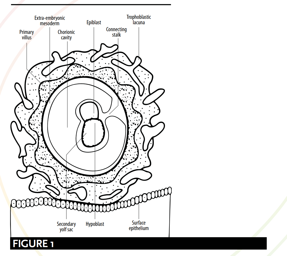
As defined by modern embryology, the myriad of meanings for the word `alaqah correspond to various stages of the embryo’s development. Comparisons can be drawn between qu’ranic and scientific depictions of the embryo's appearance and its relationship with the womb. According to modern embryology, from day 15 the embryo is hanging or suspended via the ‘connecting stalk’ [see Figure 1] and it obtains nutrients through contact with the maternal blood vessels. This description bears a striking resemblance to the picture painted by the word `alaqah – a ‘hanging’ or ‘suspended’ substance, obtaining nutrients from its host’s blood.
Im Anschluss daran finden in der 4. Woche zwei Prozesse statt: die Bildung des Gehirns und des Rückenmarks, bekannt als Neurulation, sowie die ersten Stadien der Faltung des Embryos. Mit dem Abschluss dieser beiden Prozesse ähnelt der Embryo einer blutegelartigen Form, auf die das Wort ʿalaqah hindeutet – eine kriechende, blutegel- oder wurmartige Substanz [siehe Abbildung 2, 3 und 4].
Following this, during the 4th week, two processes occur: the formation of the brain and the spinal cord, known as neurulation; and the initial stages of the folding of the embryo. It is upon the culmination of these two processes that the embryo resembles a leech-like form suggested by the word `alaqah – a creeping, leech or worm-like substance [see Figure 2, 3 and 4].
HÄNGEN/AUFGEHÄNGT
HANGING/SUSPENDED
Die Embryologen Barry Mitchell und Ram Sharma erläutern die „hängenden“ bzw. „schwebenden“ Aspekte der ʿAlaqah-Phase. Sie beschreiben den Embryo als „durch einen Verbindungsstiel aus extraembryonalem Mesoderm (primitives Bindegewebe) mit dem Zytotrophoblasten verbunden. Der Stiel ist der Vorläufer der Nabelschnur.“ 50 Abbildung 1 zeigt den Embryo, der über einen Verbindungsstiel mit dem Zytotrophoblasten verbunden ist, als würde er hängen oder schweben.
Embryologists Barry Mitchell and Ram Sharma explain the ‘hanging’ or ‘suspended’ aspects of the `alaqah stage. They describe the embryo as being, “connected to the cytotrophoblast by a connecting stalk of extra-embryonic mesoderm (primitive connective tissue). The stalk is the forerunner of the umbilical cord.” 50 Figure 1 shows the embryo connected to the cytotrophoblast via a connecting stalk, as if it were hanging or suspended.
NÄHRSTOFFZUFUHR ÜBER DIE BLUTGEFÄSSE
OBTAINING NUTRIENTS VIA BLOOD VESSELS
Im Hinblick auf die Art und Weise, wie der Embryo seine Nährstoffe durch den Kontakt mit den mütterlichen Blutgefäßen erhält, schreiben Barry Mitchell und Ram Sharma: Aufgrund des raschen Wachstums des Embryos während der zweiten Woche besteht ein Bedarf an einem effizienteren Mittel für den Nährstoff- und Gasaustausch. Dies wird erreicht, wenn die embryonalen Blutgefäße des Chorions mit den mütterlichen Blutgefäßen der Decidua in Kontakt kommen.51.
With regard to the way the embryo obtains its nutrients through contact with the maternal blood vessels, Barry Mitchell and Ram Sharma write: Due to the rapid growth of the embryo during the second week, there is a need for a more efficient means of nutritional and gaseous exchange. This is achieved when the embryonic blood vessels of the chorion come into contact with the maternal blood vessels of the decidua.51.
... [in der dritten Woche]... Der Austausch von Nährstoffen, Atemgasen und Stoffwechselabfallprodukten zwischen dem mütterlichen und dem fetalen Blut erfolgt über die plazentare Membran innerhalb der intervillösen Räume. Mütterliches Blut gelangt aus den Spiralarterien, Ästen der Uterusarterie, in diese Räume und bringt Nährstoffe und Sauerstoff für den Embryo und den Fötus.52
... [by the third week]... The exchange of nutrients, respiratory gases and waste products between the maternal and fetal blood takes place across the placental membrane within intervillous spaces. Maternal blood enters these spaces from the spiral arteries, branches of the uterine artery, bringing nutrients and oxygen for the embryo and fetus.52
Dies bekräftigt die Gültigkeit der Bedeutungen von ʿAlaqah und ruft Bilder eines Wesens hervor, das seine Nährstoffe über das Blut erhält.
This reinforces the validity of the meanings of `alaqah, and its evoking images of an entity obtaining its nutrients via blood.
NEURULATION UND DIE FALTUNG DES EMBRYOS
NEURULATION & THE FOLDING OF THE EMBRYO
Das Stadium der ʿAlaqah deutet auf den Prozess der Neurulation und die ersten Stadien der Faltung des Embryos hin. Die Neurulation umfasst die Bildung des Gehirns und des Rückenmarks etwa vom 19. bis zum 25. Tag, und die Faltung des Embryos bewirkt, dass Kopf und Schwanz einander näherkommen. Die Kombination dieser beiden physiologischen Veränderungen führt dazu, dass der Embryo einer blutegelartigen Substanz ähnelt.
The `alaqah stage suggests the process of neurulation and the initial stages of the folding of the embryo. Neurulation comprises of the formation of the brain and the spinal cord from days 19 to 25 (approx.); and the folding of the embryo involves the head and the tail being brought closer together. The combination of these two physiological changes causes the embryo to resemble a leech-like substance.
NEURULATION
NEURULATION
Am Ende der Neurulation schließen sich das kraniale und das kaudale Ende des Neuralrohrs. Der Embryo erhält zu diesem Zeitpunkt ein blutegelartiges Aussehen 53 [siehe Abbildung 2].
At the end of neurulation the cranial and caudal ends of the neural tube close. The embryo, at this point, becomes leech-like in appearance 53 [see Figure 2].
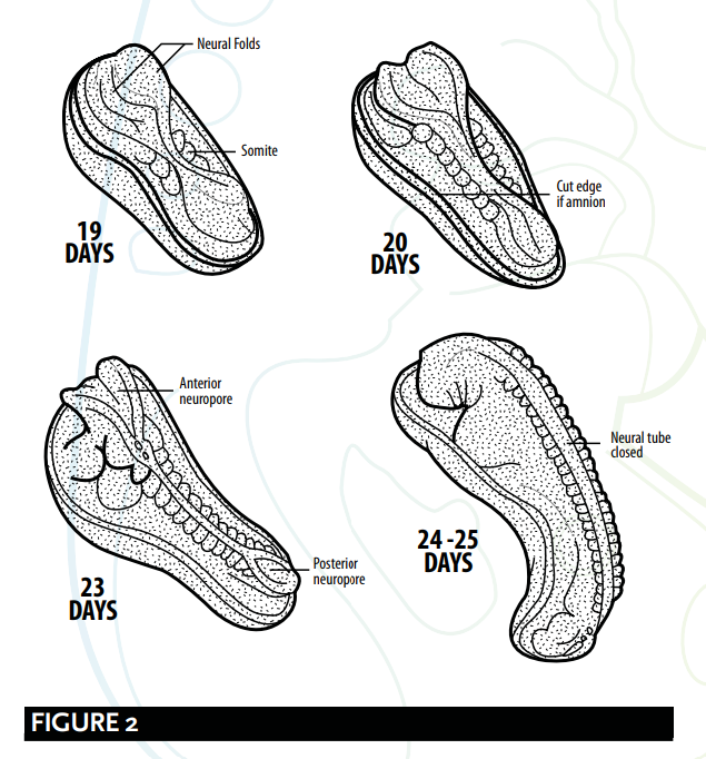
Barry Micthell und Ram Sharma erklären den Prozess der Neurulation.
Barry Micthell and Ram Sharma explain the process of neurulation.
Im cranialen Ende des Primitivstreifens induzieren etwa am 19. Tag das darunterliegende Mesoderm und die Chorda dorsalis das Ektoderm zur Bildung der Neuralplatte, die sich zu den Neuralwülsten aufwölbt. Die Neuralplatte vergrößert sich zunächst am kranialen Ende. Am 20. Tag bleibt die Neuralplatte im mittleren Bereich des Embryos schmal, dehnt sich jedoch am kaudalen Ende aus. Die Platte vertieft sich zur Neuralrinne, aus der sich das Neuralrohr bildet. Das kraniale und das kaudale Ende des Rohrs sind offen und werden als vorderer bzw. hinterer Neuroporus bezeichnet; diese schließen sich schließlich. 54
At about 19 days, at the cranial end of the primitive streak, the underlying mesoderm and notochord induce the ectoderm to form the neural plate, which rounds up to form the neural folds. The neural plate enlarges initially at the cranial end. At 20 days, the neural plate in the mid-region of the embryo remains narrowed, but it expands at the caudal end. The plate deepens to form the neural groove from which the neural tube forms. The cranial and caudal ends of the tube are open and are known as the anterior and posterior neuropores; these eventually close. 54
FALTUNG DES EMBRYOS
FOLDING OF THE EMBRYO
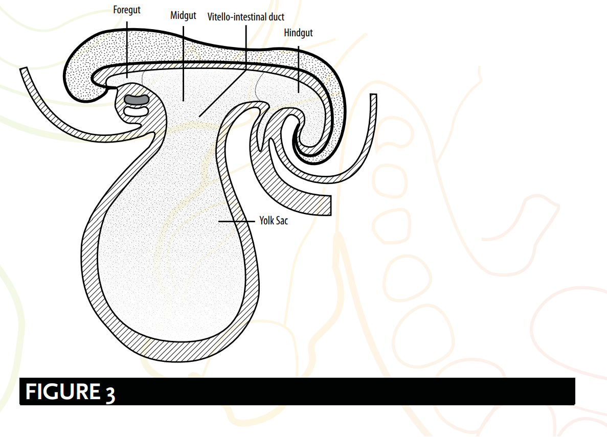
Die Faltung des Embryos ist außerdem dafür verantwortlich, dass eine blutegelartige Gestalt entsteht [siehe Abbildung 3] – oder, wie Embryologen es beschrieben haben, eine zylindrische bzw. röhrenartige Struktur. Die Embryologen Keith Moore und T. V. N. Persaud legen nahe, dass:
The folding of the embryo is also responsible for forming a leech-like shape [see Figure 3], or as embryologists have described; a cylindric or tube-like structure. Embryologists Keith Moore and T. V. N. Persaud suggest that:
Ein bedeutendes Ereignis bei der Ausbildung der Körperform ist die Faltung der flachen trilaminaren Keimscheibe zu einem annähernd zylindrischen Embryo. Die Faltung resultiert aus dem raschen Wachstum des Embryos, insbesondere des Gehirns und des Rückenmarks. 55
A significant event in the establishment of body form is the folding of the flat trilaminar embryonic disc into a somewhat cylindric embryo. Folding results from the rapid growth of the embryo, particularly the brain and the spinal cord. 55
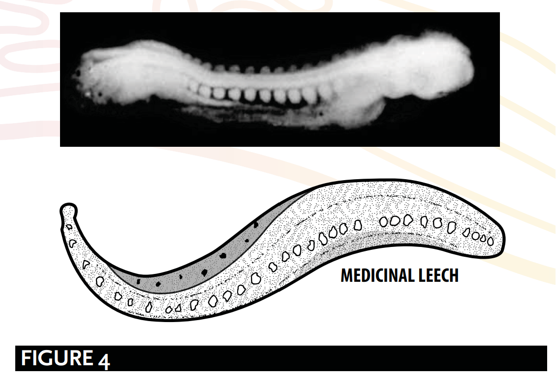
Die röhren- oder egelartige Struktur [siehe Abbildung 4], wie sie von Barry Mitchell und Ram Sharma erklärt wird, ist bedingt durch:
The tube-like or leech-like structure [see Figure 4], as explained by Barry Mitchell and Ram Sharma, is due to:
Die Längsfaltung, die zwischen dem 21. und 24. Tag stattfindet, führt dazu, dass sich der Embryo so krümmt, dass Kopf und Schwanz einander näher gebracht werden, … um eine röhrenartige Struktur zu bilden. 56
longitudinal folding, which occurs between days 21 and 24, result[ing] in … the embryo [bending] so that the head and tail are brought closer together...[to] form a tube-like structure. 56
ZUSAMMENFASSUNG
SUMMARY
Die Phase der ʿAlaqa:
The `alaqah stage:
Der Embryo ist über einen Verbindungsstiel mit dem Zytotrophoblasten verbunden, so als würde er hängen oder schweben.
The embryo is connected to the cytotrophoblast via a connecting stalk, as if it were hanging or suspended.
Die Bildung des Gehirns und des Rückenmarks, bekannt als Neurulation, sowie während der anfänglichen Stadien der Faltung des Embryos. Mit dem Abschluss dieser Vorgänge erscheint der Embryo blutegelartig.
The formation of the brain and the spinal cord, known as neurulation; and during the initial stages of the folding of the embryo. It is upon the culmination of these processes that the embryo appears leech-like.
Der Embryo bezieht seine Nährstoffe über den Kontakt mit den Blutgefäßen der Mutter. Dies gleicht der Wirkungsweise einer blutsaugerähnlichen Substanz, die ihre Nährstoffe über das Blut bezieht.
The embryo obtains its nutrients via contact with the maternal blood vessels. This mirrors the action of a leech-like substance obtaining its nutrients via blood.
GALEN & ʿALAQAH
GALEN & `ALAQAH
Kritiker behaupten, dass die Verwendung des Wortes ʿalaqah eine Beschreibung der zweiten Stufe des menschlichen Embryos nach Galen sei. Ihre Übersetzung des Wortes ʿalaqah als „Blutgerinnsel“ erwecke den Eindruck, es handle sich um Galens Beschreibung dieser Stufe.
Critics declare that the use of the word `alaqah is a description of Galen’s second stage of the human embryo. Their translation of the word `alaqah to ‘blood clot’ creates an impression of Galen's description of this stage:
Doch wenn sie [der Blutklumpen] mit Blut gefüllt ist und Herz, Gehirn und Leber noch nicht gegliedert und nicht geformt sind, aber inzwischen eine gewisse Festigkeit und beträchtliche Größe haben, dann ist dies die zweite Phase; die Substanz des Fötus hat nun die Form von Fleisch und nicht mehr die Form von Samen. 57
But when it [the clot] has been filled with blood, and heart, brain and liver are still unarticulated and unshaped yet have by now certain solidarity and considerable size, this is the second period; the substance of the foetus has the form of flesh and no longer the form of semen. 57
Es gibt eine Reihe von Schwächen in den Implikationen dieser Kritiker:
There are a number of weaknesses with the implications of these critics:
1. Erstens erwähnt der Qur’an nicht das „Herz, Gehirn und Leber...
1. Firstly, the Qur’an does not make a mention of the “heart, brain and liver...
„[Dies war] noch unartikuliert und ungeformt, doch mit einer gewissen Solidarität und von beträchtlicher Größe“.
[being] unarticulated and unshaped yet [with] a certain solidarity and [of a] considerable size”.
2. Zweitens bedeutet das Wort ʿalaqah nicht Blut; vielmehr sind seine primären Bedeutungen „eine sich anklammernde Gestalt“ und „ein blutegelartiger Stoff“. Einige arabische Sprachwissenschaftler schreiben dem Wort ʿalaqah die Bedeutung „Blutgerinnsel“ zu, nicht weil es mit Blut gefüllt ist, sondern aufgrund seines Wesens des „Anklammerns“. Diese Deutung entspricht der primären Bedeutung des Wortes.
2. Secondly, the word `alaqah does not mean blood; rather, its primary meanings are ‘a clinging form’ and ‘a leech-like substance’. Some Arab linguists would attribute the meaning of a ‘blood clot’ to the word `alaqah, not because of it being filled with blood, but instead due to it ‘clinging’ by nature. This interpretation matches the primary meaning of the word.
3. Drittens besteht Galen darauf, dass der Fötus in der zweiten Phase „die Form von Fleisch hat“, während der Qur'an bestätigt, dass die Phase der mudghah (ein gekautes Stück Fleisch oder ein Fleischklumpen) auf die Phase der ʿalaqah folgt.
3. Thirdly, Galen insists “the foetus has the form of flesh” during the second stage, whereas the Qur’an affirms the mudghah (a chewed piece of meat or lump of flesh) stage comes after the `alaqah stage.
4. Viertens schreibt Galen in seinem Buch Über die natürlichen Fähigkeiten: So verhält es sich auch mit dem Samen: Seine Fähigkeiten besaß er von Anfang an.58 Galens unzutreffende Schlussfolgerung, der Samen allein sei für das genetische Material verantwortlich, ist mit dem Koran und den prophetischen Überlieferungen unvereinbar und steht im direkten Widerspruch zu ihnen. Wie bereits erörtert (siehe Aristoteles, Galen & Nutfah), betrachten die islamischen Quellentexte die Nutfah als einen vermischten Tropfen oder eine Flüssigkeit sowohl vom Mann als auch von der Frau, und sie bestätigen, dass sowohl der Mann als auch die Frau für die Genetik des Kindes verantwortlich sind.59 Wenn der Prophet ﷺ also Galens Ansichten über die menschliche Entwicklung übernommen hätte, wie kommt es dann, dass er die unzutreffende Wissenschaft verwarf und nur das verwendete, was richtig war, obwohl man im Arabien des 7. Jahrhunderts keine Möglichkeit hatte, zu wissen, welche Ansichten zutreffend waren?
FALSCHDARSTELLUNG DES AUSSEHENS DES EMBRYOS
MISREPRESENTING THE EMBRYO’S APPEARANCE
Zeitgenössische Kommentatoren argumentieren, dass der Embryo nur dann blutegelartig aussieht, wenn der Dottersack entfernt wird, und dass die Beschreibung des ʿAlaqah daher eine Falschdarstellung des Aussehens des Embryos in diesem Stadium sei. Embryologen erklären jedoch, dass der Dottersack durch ein außerembryonales Merkmal, das sogenannte extraembryonale Coelom, vom Embryo getrennt ist. Diese Tatsache widerlegt Behauptungen einer Falschdarstellung, da der Dottersack nicht Teil der Struktur des Embryos ist.60
Contemporary commentators argue the embryo only looks leech-like when the yolk sac is removed, and that the `alaqah's description is therefore a misrepresentation of the embryo’s appearance at this stage. However, embryologists explain that the yolk sac is separated from the embryo by an extra-embryonic feature, called the extra-embryonic coelom. This fact disproves claims of misrepresentation as the yolk sac is not part of the structure of the embryo.60