4. Une forme adhérente
4. A Clinging Form
Puis Nous avons fait de cette goutte une adhérence
Then We made that drop of fluid into a clinging form
Le Coran décrit l'étape suivante du développement de l'embryon humain avec le mot `alaqah. Ce mot revêt diverses significations, notamment : pendre, être suspendu, être ballant, coller, s'accrocher, se fendre et adhérer. Il peut également signifier attraper, être capturé, être fixé ou être joint.48
The Qur’an describes the next stage of the developing human embryo with the word `alaqah. This word carries various meanings including: to hang, to be suspended, to be dangled, to stick, to cling, to cleave and to adhere. It can also mean to catch, to get caught, to be affixed or subjoined.48
D'autres connotations du mot `alaqah incluent une substance semblable à une sangsue, ayant la ressemblance d'un ver ; ou ayant une disposition « rampante » inclinée à sucer le sang. Enfin, son sens inclut l'argile qui adhère à la main et le sang épais et coagulé - en raison de son adhérence. 49
Other connotations of the word `alaqah include a leech-like substance, having the resemblance of a worm; or being of a ‘creeping’ disposition inclined to the sucking of blood. Finally, its meaning includes clay that clings to the hand and thick, clotted blood - because of its clinging together. 49
INTERPRÉTATION SCIENTIFIQUE
SCIENTIFIC INTERPRETATION
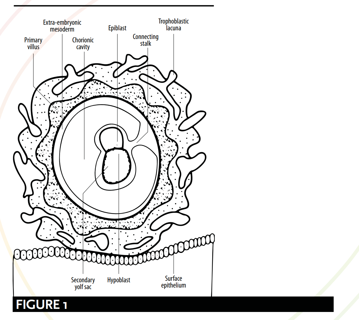
Telle que définie par l'embryologie moderne, la myriade de significations du mot `alaqah correspond aux diverses étapes du développement de l'embryon. Des comparaisons peuvent être établies entre les descriptions coraniques et scientifiques de l'apparence de l'embryon et de sa relation avec l'utérus. Selon l'embryologie moderne, à partir du 15ème jour, l'embryon est suspendu via le « pédicule de connexion » [voir Figure 1] et il obtient des nutriments par contact avec les vaisseaux sanguins maternels. Cette description présente une ressemblance frappante avec l'image dépeinte par le mot `alaqah – une substance « suspendue » ou « accrochée », obtenant des nutriments du sang de son hôte.
As defined by modern embryology, the myriad of meanings for the word `alaqah correspond to various stages of the embryo’s development. Comparisons can be drawn between qu’ranic and scientific depictions of the embryo's appearance and its relationship with the womb. According to modern embryology, from day 15 the embryo is hanging or suspended via the ‘connecting stalk’ [see Figure 1] and it obtains nutrients through contact with the maternal blood vessels. This description bears a striking resemblance to the picture painted by the word `alaqah – a ‘hanging’ or ‘suspended’ substance, obtaining nutrients from its host’s blood.
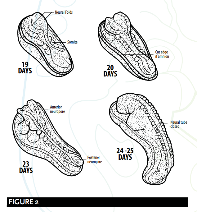
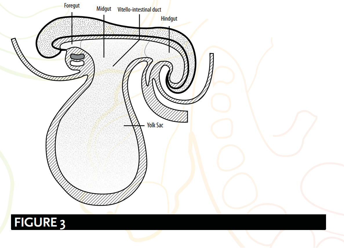
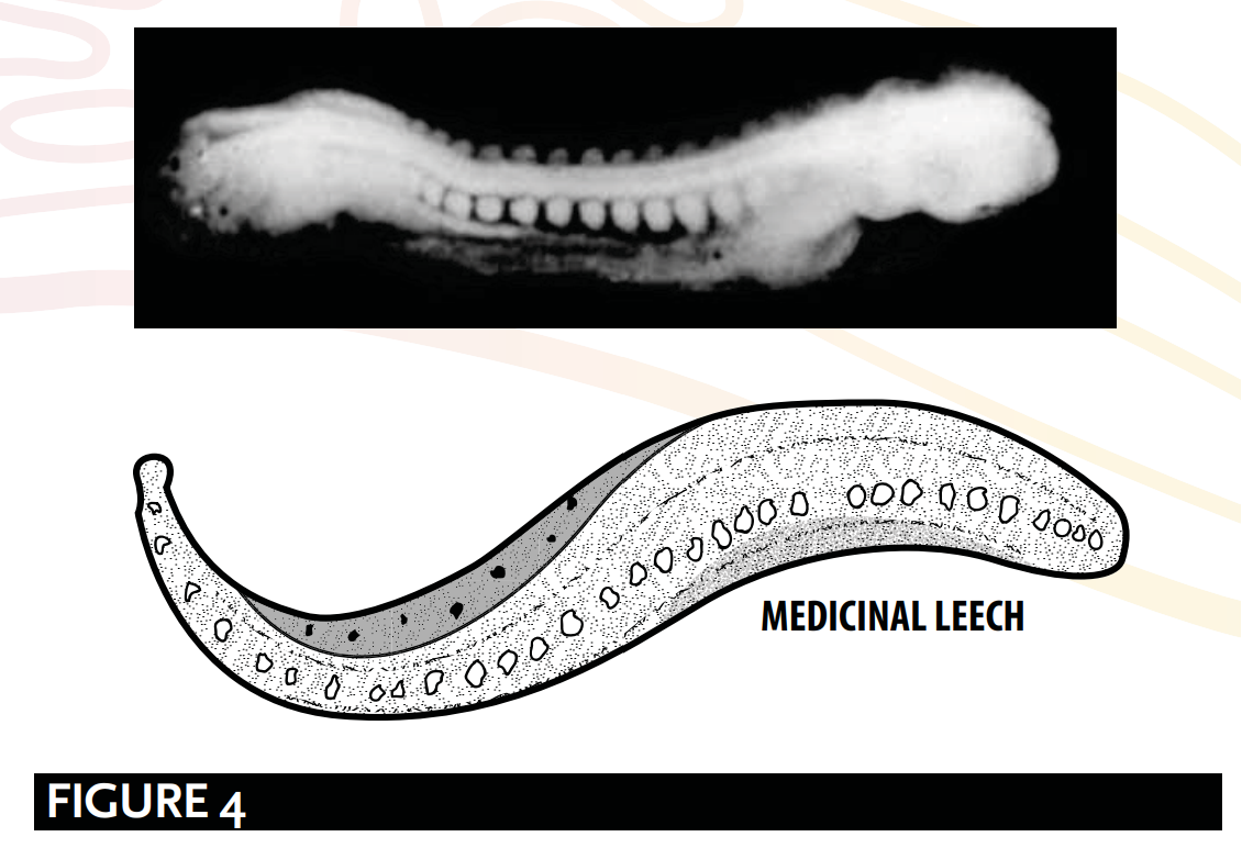
À la suite de cela, durant la 4ème semaine, deux processus se produisent : la formation du cerveau et de la moelle épinière, connue sous le nom de neurulation ; et les premières étapes du repliement de l'embryon. C'est à l'aboutissement de ces deux processus que l'embryon ressemble à une forme de sangsue suggérée par le mot `alaqah – une substance rampante, semblable à une sangsue ou à un ver [voir Figure 2, 3 et 4].
Following this, during the 4th week, two processes occur: the formation of the brain and the spinal cord, known as neurulation; and the initial stages of the folding of the embryo. It is upon the culmination of these two processes that the embryo resembles a leech-like form suggested by the word `alaqah – a creeping, leech or worm-like substance [see Figure 2, 3 and 4].
PENDANT/SUSPENDU
HANGING/SUSPENDED
Les embryologistes Barry Mitchell et Ram Sharma expliquent les aspects « suspendus » de l'étape de l' `alaqah. Ils décrivent l'embryon comme étant « connecté au cytotrophoblaste par un pédicule de connexion de mésoderme extra-embryonnaire (tissu conjonctif primitif). Le pédicule est le précurseur du cordon ombilical. » 50 La Figure 1 montre l'embryon connecté au cytotrophoblaste via un pédicule de connexion, comme s'il était suspendu.
Embryologists Barry Mitchell and Ram Sharma explain the ‘hanging’ or ‘suspended’ aspects of the `alaqah stage. They describe the embryo as being, “connected to the cytotrophoblast by a connecting stalk of extra-embryonic mesoderm (primitive connective tissue). The stalk is the forerunner of the umbilical cord.” 50 Figure 1 shows the embryo connected to the cytotrophoblast via a connecting stalk, as if it were hanging or suspended.
L'OBTENTION DES NUTRIMENTS VIA LES VAISSEAUX SANGUINS
OBTAINING NUTRIENTS VIA BLOOD VESSELS
Concernant la manière dont l'embryon obtient ses nutriments par contact avec les vaisseaux sanguins maternels, Barry Mitchell et Ram Sharma écrivent : En raison de la croissance rapide de l'embryon durant la deuxième semaine, il est nécessaire de disposer d'un moyen plus efficace d'échange nutritionnel et gazeux. Cela est réalisé lorsque les vaisseaux sanguins embryonnaires du chorion entrent en contact avec les vaisseaux sanguins maternels de la décidue.51.
With regard to the way the embryo obtains its nutrients through contact with the maternal blood vessels, Barry Mitchell and Ram Sharma write: Due to the rapid growth of the embryo during the second week, there is a need for a more efficient means of nutritional and gaseous exchange. This is achieved when the embryonic blood vessels of the chorion come into contact with the maternal blood vessels of the decidua.51.
... [dès la troisième semaine]... L'échange de nutriments, de gaz respiratoires et de déchets entre le sang maternel et le sang fœtal s'effectue à travers la membrane placentaire au sein des espaces intervillosités. Le sang maternel pénètre dans ces espaces par les artères spiralées, branches de l'artère utérine, apportant les nutriments et l'oxygène pour l'embryon et le fœtus.52
... [by the third week]... The exchange of nutrients, respiratory gases and waste products between the maternal and fetal blood takes place across the placental membrane within intervillous spaces. Maternal blood enters these spaces from the spiral arteries, branches of the uterine artery, bringing nutrients and oxygen for the embryo and fetus.52
Ceci renforce la validité des significations de `alaqah, et l'évocation d'images d'une entité tirant ses nutriments via le sang.
This reinforces the validity of the meanings of `alaqah, and its evoking images of an entity obtaining its nutrients via blood.
NEURULATION ET LE PLIAGE DE L'EMBRYON
NEURULATION & THE FOLDING OF THE EMBRYO
L'étape de l' `alaqah suggère le processus de la neurulation et les premières étapes du repliement de l'embryon. La neurulation comprend la formation du cerveau et de la moelle épinière du 19e au 25e jour environ ; et le repliement de l'embryon implique que la tête et la queue se rapprochent l'une de l'autre. La combinaison de ces deux changements physiologiques fait que l'embryon ressemble à une substance semblable à une sangsue.
The `alaqah stage suggests the process of neurulation and the initial stages of the folding of the embryo. Neurulation comprises of the formation of the brain and the spinal cord from days 19 to 25 (approx.); and the folding of the embryo involves the head and the tail being brought closer together. The combination of these two physiological changes causes the embryo to resemble a leech-like substance.
NEURULATION
NEURULATION
À la fin de la neurulation, les extrémités crânienne et caudale du tube neural se ferment. À ce stade, l'embryon prend une apparence semblable à celle d'une sangsue 53 [voir Figure 2].
At the end of neurulation the cranial and caudal ends of the neural tube close. The embryo, at this point, becomes leech-like in appearance 53 [see Figure 2].
Barry Micthell et Ram Sharma expliquent le processus de la neurulation.
Barry Micthell and Ram Sharma explain the process of neurulation.
Vers le 19ème jour, à l'extrémité crâniale de la ligne primitive, le mésoderme sous-jacent et la chorde induisent l'ectoderme à former la plaque neurale, qui s'arrondit pour former les replis neuraux. La plaque neurale s'élargit initialement à l'extrémité crâniale. À 20 jours, la plaque neurale dans la région médiane de l'embryon reste étroite, mais elle s'étend à l'extrémité caudale. La plaque s'approfondit pour former le sillon neural à partir duquel le tube neural se forme. Les extrémités crâniale et caudale du tube sont ouvertes et sont connues sous le nom de neuropores antérieur et postérieur ; celles-ci finissent par se fermer. 54
At about 19 days, at the cranial end of the primitive streak, the underlying mesoderm and notochord induce the ectoderm to form the neural plate, which rounds up to form the neural folds. The neural plate enlarges initially at the cranial end. At 20 days, the neural plate in the mid-region of the embryo remains narrowed, but it expands at the caudal end. The plate deepens to form the neural groove from which the neural tube forms. The cranial and caudal ends of the tube are open and are known as the anterior and posterior neuropores; these eventually close. 54
PLIAGE DE L'EMBRYON
FOLDING OF THE EMBRYO
Le repliement de l'embryon est également responsable de la formation d'une forme semblable à une sangsue [voir Figure 3], ou comme l'ont décrit les embryologistes : une structure cylindrique ou en forme de tube. Les embryologistes Keith Moore et T. V. N. Persaud suggèrent que :
The folding of the embryo is also responsible for forming a leech-like shape [see Figure 3], or as embryologists have described; a cylindric or tube-like structure. Embryologists Keith Moore and T. V. N. Persaud suggest that:
Un événement significatif dans l'établissement de la forme corporelle est le repliement du disque embryonnaire trilaminaire plat en un embryon quelque peu cylindrique. Le repliement résulte de la croissance rapide de l'embryon, particulièrement du cerveau et de la moelle épinière. 55
A significant event in the establishment of body form is the folding of the flat trilaminar embryonic disc into a somewhat cylindric embryo. Folding results from the rapid growth of the embryo, particularly the brain and the spinal cord. 55
La structure en forme de tube ou de sangsue [voir Figure 4], telle qu'expliquée par Barry Mitchell et Ram Sharma, est due à :
The tube-like or leech-like structure [see Figure 4], as explained by Barry Mitchell and Ram Sharma, is due to:
le repliement longitudinal, qui se produit entre les 21e et 24e jours, entraîne... la courbure de l'embryon de sorte que la tête et la queue se rapprochent... pour former une structure tubulaire. 56
longitudinal folding, which occurs between days 21 and 24, result[ing] in … the embryo [bending] so that the head and tail are brought closer together...[to] form a tube-like structure. 56
RÉSUMÉ
SUMMARY
L'étape de l'alaqah :
The `alaqah stage:
L'embryon est relié au cytotrophoblaste par un pédicule de connexion, comme s'il était suspendu ou en lévitation.
The embryo is connected to the cytotrophoblast via a connecting stalk, as if it were hanging or suspended.
La formation du cerveau et de la moelle épinière, connue sous le nom de neurulation ; et durant les premières étapes du repliement de l'embryon. C'est au terme de ces processus que l'embryon prend une apparence semblable à celle d'une sangsue.
The formation of the brain and the spinal cord, known as neurulation; and during the initial stages of the folding of the embryo. It is upon the culmination of these processes that the embryo appears leech-like.
L'embryon obtient ses nutriments par contact avec les vaisseaux sanguins maternels. Cela reflète l'action d'une substance semblable à une sangsue qui obtient ses nutriments par le sang.
The embryo obtains its nutrients via contact with the maternal blood vessels. This mirrors the action of a leech-like substance obtaining its nutrients via blood.
GALEN & `ALAQAH
GALEN & `ALAQAH
Les critiques affirment que l'utilisation du mot `alaqah est une description de la deuxième étape de l'embryon humain selon Galien. Leur traduction du mot `alaqah par « caillot de sang » crée une impression de la description de Galien de cette étape :
Critics declare that the use of the word `alaqah is a description of Galen’s second stage of the human embryo. Their translation of the word `alaqah to ‘blood clot’ creates an impression of Galen's description of this stage:
Mais lorsqu'il [l'caillot] s'est rempli de sang, et que le cœur, le cerveau et le foie ne sont pas encore articulés ni formés, mais possèdent désormais une certaine solidarité et une taille considérable, c'est la deuxième période ; la substance du fœtus a la forme de chair et non plus la forme de sperme. 57
But when it [the clot] has been filled with blood, and heart, brain and liver are still unarticulated and unshaped yet have by now certain solidarity and considerable size, this is the second period; the substance of the foetus has the form of flesh and no longer the form of semen. 57
Il existe un certain nombre de faiblesses dans les implications de ces critiques :
There are a number of weaknesses with the implications of these critics:
1. Premièrement, le Coran ne mentionne pas le « cœur, le cerveau et le foie...
1. Firstly, the Qur’an does not make a mention of the “heart, brain and liver...
« [étant] inarticulé et informe, tout en possédant une certaine solidarité et [étant d'une] taille considérable ».
[being] unarticulated and unshaped yet [with] a certain solidarity and [of a] considerable size”.
2. Deuxièmement, le mot `alaqah ne signifie pas le sang ; ses significations primaires sont plutôt « une forme adhérente » et « une substance semblable à une sangsue ». Certains linguistes arabes attribueraient le sens de « caillot de sang » au mot `alaqah, non pas parce qu'il est rempli de sang, mais plutôt en raison de sa nature « adhérente ». Cette interprétation correspond au sens primaire du mot.
2. Secondly, the word `alaqah does not mean blood; rather, its primary meanings are ‘a clinging form’ and ‘a leech-like substance’. Some Arab linguists would attribute the meaning of a ‘blood clot’ to the word `alaqah, not because of it being filled with blood, but instead due to it ‘clinging’ by nature. This interpretation matches the primary meaning of the word.
3. Troisièmement, Galen insiste sur le fait que « le fœtus a la forme de chair » durant la deuxième étape, alors que le Coran affirme que l'étape de la *mudghah* (un morceau de viande mâché ou une masse de chair) vient après l'étape de l'*alaqah*.
3. Thirdly, Galen insists “the foetus has the form of flesh” during the second stage, whereas the Qur’an affirms the mudghah (a chewed piece of meat or lump of flesh) stage comes after the `alaqah stage.
4. Quatrièmement, dans son livre *On the Natural Faculties*, Galien écrit : ainsi en est-il du sperme : ses facultés, il les possédait dès le commencement.58 L'implication incorrecte de Galien selon laquelle le sperme serait l'unique responsable du matériel génétique est incompatible, et en opposition directe, avec le Coran et les traditions prophétiques. Comme exploré précédemment (voir *Aristotle, Galen & Nutfah*), les textes sources islamiques considèrent la *nutfah* comme une goutte ou un fluide mêlé provenant à la fois de l'homme et de la femme, et confirment que l'homme et la femme sont tous deux responsables de la génétique de l'enfant.59 Par conséquent, si le Prophète ﷺ avait copié les vues de Galien sur le développement humain, comment se fait-il qu'il ait rejeté la science incorrecte pour n'utiliser que ce qui était correct, alors qu'il n'y avait aucun moyen, dans l'Arabie du VIIe siècle, de savoir quelles vues étaient exactes ?
LA REPRÉSENTATION ERRONÉE DE L'APPARENCE DE L'EMBRYON
MISREPRESENTING THE EMBRYO’S APPEARANCE
Les commentateurs contemporains soutiennent que l'embryon ne ressemble à une sangsue que lorsque le sac vitellin est retiré, et que la description de l'`alaqah est donc une représentation erronée de l'apparence de l'embryon à ce stade. Cependant, les embryologistes expliquent que le sac vitellin est séparé de l'embryon par une structure extra-embryonnaire, appelée le coelome extra-embryonnaire. Ce fait infirme les affirmations de représentation erronée, car le sac vitellin ne fait pas partie de la structure de l'embryon.60
Contemporary commentators argue the embryo only looks leech-like when the yolk sac is removed, and that the `alaqah's description is therefore a misrepresentation of the embryo’s appearance at this stage. However, embryologists explain that the yolk sac is separated from the embryo by an extra-embryonic feature, called the extra-embryonic coelom. This fact disproves claims of misrepresentation as the yolk sac is not part of the structure of the embryo.60