4. Een kleverende vorm
4. A Clinging Form
Toen maakten Wij die druppel tot een vastzittende vorm
Then We made that drop of fluid into a clinging form
De Qur'an beschrijft de volgende fase van de ontwikkelende menselijke embryo met het woord `alaqah. Dit woord draagt verschillende betekenissen, waaronder: hangen, opgeschort zijn, bungelen, plakken, kleven, splitsen en hechten. Het kan ook betekenen: vangen, gevangen worden, bevestigd zijn of worden toegevoegd.48
The Qur’an describes the next stage of the developing human embryo with the word `alaqah. This word carries various meanings including: to hang, to be suspended, to be dangled, to stick, to cling, to cleave and to adhere. It can also mean to catch, to get caught, to be affixed or subjoined.48
Andere connotaties van het woord `alaqah omvatten een aderwurm-achtige substantie, die de gelijkenis heeft van een worm; of een 'kruipende' aard hebben, geneigd tot het zuigen van bloed. Ten slotte omvat de betekenis klei die aan de hand kleeft en dik, gestold bloed - vanwege het aan elkaar kleven. 49
Other connotations of the word `alaqah include a leech-like substance, having the resemblance of a worm; or being of a ‘creeping’ disposition inclined to the sucking of blood. Finally, its meaning includes clay that clings to the hand and thick, clotted blood - because of its clinging together. 49
WETENSCHAPPELIJKE INTERPRETATIE
SCIENTIFIC INTERPRETATION
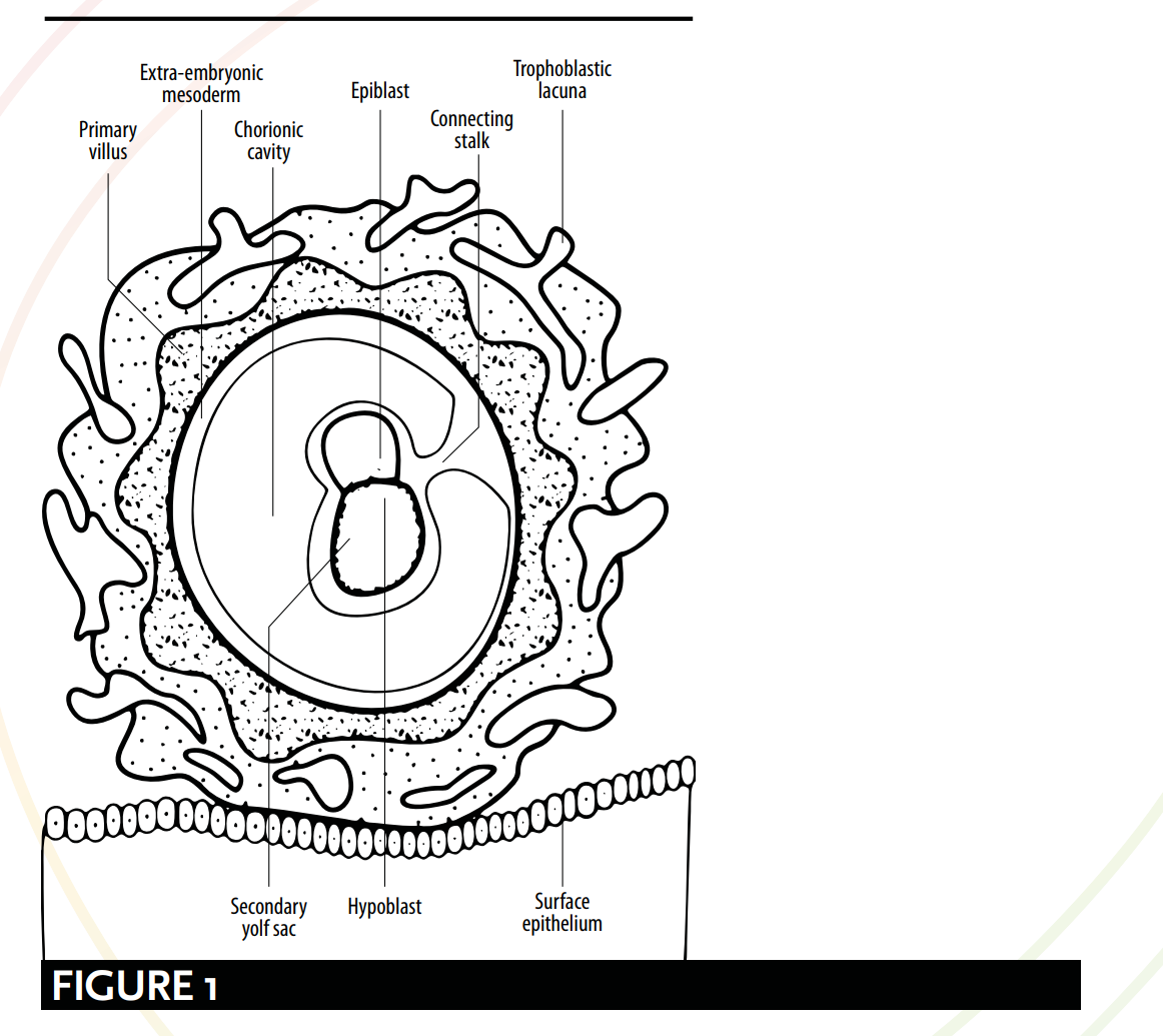
Zoals gedefinieerd door de moderne embryologie, komen de talloze betekenissen van het woord `alaqah overeen met verschillende stadia van de ontwikkeling van het embryo. Er kunnen vergelijkingen worden getrokken tussen de koranische en wetenschappelijke beschrijvingen van het uiterlijk van het embryo en de relatie ervan met de baarmoeder. Volgens de moderne embryologie hangt het embryo vanaf dag 15 of is het opgehangen via de ‘verbindingssteel’ [zie Figuur 1] en verkrijgt het voedingsstoffen door contact met de moederlijke bloedvaten. Deze beschrijving vertoont een opvallende gelijkenis met het beeld dat geschetst wordt door het woord `alaqah – een ‘hangende’ of ‘opgehangen’ substantie, die voedingsstoffen verkrijgt uit het bloed van zijn gastheer.
As defined by modern embryology, the myriad of meanings for the word `alaqah correspond to various stages of the embryo’s development. Comparisons can be drawn between qu’ranic and scientific depictions of the embryo's appearance and its relationship with the womb. According to modern embryology, from day 15 the embryo is hanging or suspended via the ‘connecting stalk’ [see Figure 1] and it obtains nutrients through contact with the maternal blood vessels. This description bears a striking resemblance to the picture painted by the word `alaqah – a ‘hanging’ or ‘suspended’ substance, obtaining nutrients from its host’s blood.
Hierna vinden tijdens de 4e week twee processen plaats: de vorming van de hersenen en het ruggenmerg, bekend als neurulatie; en de beginfases van het vouwen van het embryo. Het is bij de voltooiing van deze twee processen dat het embryo een bloedzuigerachtige vorm aanneemt, gesuggereerd door het woord `alaqah – een kruipende, bloedzuiger- of wormachtige substantie [zie Figuur 2, 3 en 4].
Following this, during the 4th week, two processes occur: the formation of the brain and the spinal cord, known as neurulation; and the initial stages of the folding of the embryo. It is upon the culmination of these two processes that the embryo resembles a leech-like form suggested by the word `alaqah – a creeping, leech or worm-like substance [see Figure 2, 3 and 4].
HANGEND/OPGEHANGEN
HANGING/SUSPENDED
Embryologen Barry Mitchell en Ram Sharma leggen de ‘hangende’ of ‘gesuspendeerde’ aspecten van de `alaqah-fase uit. Zij beschrijven het embryo als zijnde, “verbonden met de cytotrofoblast door een verbindingssteel van extra-embryonale mesoderm (primitief bindweefsel). De steel is de voorloper van de navelstreng.” 50 Figuur 1 laat het embryo zien dat via een verbindingssteel met de cytotrofoblast is verbonden, alsof het hangt of gesuspendeerd is.
Embryologists Barry Mitchell and Ram Sharma explain the ‘hanging’ or ‘suspended’ aspects of the `alaqah stage. They describe the embryo as being, “connected to the cytotrophoblast by a connecting stalk of extra-embryonic mesoderm (primitive connective tissue). The stalk is the forerunner of the umbilical cord.” 50 Figure 1 shows the embryo connected to the cytotrophoblast via a connecting stalk, as if it were hanging or suspended.
HET BEMETIGEN VAN VOEDINGSSTOFFEN VIA BLOEDVATAAN
OBTAINING NUTRIENTS VIA BLOOD VESSELS
Met betrekking tot de wijze waarop de embryo zijn voedingsstoffen verkrijgt door contact met de moederlijke bloedvaten, schrijven Barry Mitchell en Ram Sharma: Vanwege de snelle groei van de embryo tijdens de tweede week is er behoefte aan een efficiëntere manier van nutritionele en gasvormige uitwisseling. Dit wordt bereikt wanneer de embryonale bloedvaten van de chorion in contact komen met de moederlijke bloedvaten van de decidua.51.
With regard to the way the embryo obtains its nutrients through contact with the maternal blood vessels, Barry Mitchell and Ram Sharma write: Due to the rapid growth of the embryo during the second week, there is a need for a more efficient means of nutritional and gaseous exchange. This is achieved when the embryonic blood vessels of the chorion come into contact with the maternal blood vessels of the decidua.51.
... [tegen de derde week]... De uitwisseling van voedingsstoffen, ademhalingsgassen en afvalstoffen tussen het moederlijke en foetale bloed vindt plaats via het placentamembraan binnen de intervilloze ruimtes. Moederlijk bloed stroomt deze ruimtes binnen vanuit de spiraalarteriën, vertakkingen van de arteria uterina, waarbij het voedingsstoffen en zuurstof voor het embryo en de foetus aanvoert.52
... [by the third week]... The exchange of nutrients, respiratory gases and waste products between the maternal and fetal blood takes place across the placental membrane within intervillous spaces. Maternal blood enters these spaces from the spiral arteries, branches of the uterine artery, bringing nutrients and oxygen for the embryo and fetus.52
Dit versterkt de geldigheid van de betekenissen van `alaqah, en het oproepen van beelden van een entiteit die zijn voedingsstoffen via bloed verkrijgt.
This reinforces the validity of the meanings of `alaqah, and its evoking images of an entity obtaining its nutrients via blood.
NEURULATIE & HET VOUWEN VAN HET EMBRYO
NEURULATION & THE FOLDING OF THE EMBRYO
De `alaqah-fase suggereert het proces van neurulatie en de beginfase van het vouwen van de embryo. Neurulatie omvat de vorming van de hersenen en het ruggenmerg van dag 19 tot 25 (ongeveer); en het vouwen van de embryo houdt in dat de kop en de staart dichter bij elkaar worden gebracht. De combinatie van deze twee fysiologische veranderingen zorgt ervoor dat de embryo lijkt op een aderwurm-achtige substantie.
The `alaqah stage suggests the process of neurulation and the initial stages of the folding of the embryo. Neurulation comprises of the formation of the brain and the spinal cord from days 19 to 25 (approx.); and the folding of the embryo involves the head and the tail being brought closer together. The combination of these two physiological changes causes the embryo to resemble a leech-like substance.
NEURULATIE
NEURULATION
Aan het einde van de neurulatie sluiten de craniale en caudale uiteinden van de neurale buis. Op dit punt krijgt het embryo een uiterlijk dat lijkt op een bloedzuiger 53 [zie Figuur 2].
At the end of neurulation the cranial and caudal ends of the neural tube close. The embryo, at this point, becomes leech-like in appearance 53 [see Figure 2].
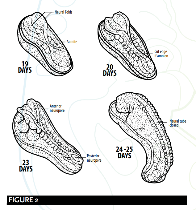
Barry Micthell en Ram Sharma leggen het proces van neurulatie uit.
Barry Micthell and Ram Sharma explain the process of neurulation.
Na ongeveer 19 dagen, aan het craniale uiteinde van de primitieve streep, induceren het onderliggende mesoderm en de notochord het ectoderm om de neurale plaat te vormen, die zich oprolt om de neurale plooien te vormen. De neurale plaat vergroot zich aanvankelijk aan het craniale uiteinde. Na 20 dagen blijft de neurale plaat in de middenregio van het embryo vernauwd, maar breidt deze zich uit aan het caudale uiteinde. De plaat verdiept zich om de neurale groef te vormen, waaruit de neurale buis ontstaat. De craniale en caudale uiteinden van de buis zijn open en staan bekend als de anterieure en posterieure neuroporen; deze sluiten uiteindelijk. 54
At about 19 days, at the cranial end of the primitive streak, the underlying mesoderm and notochord induce the ectoderm to form the neural plate, which rounds up to form the neural folds. The neural plate enlarges initially at the cranial end. At 20 days, the neural plate in the mid-region of the embryo remains narrowed, but it expands at the caudal end. The plate deepens to form the neural groove from which the neural tube forms. The cranial and caudal ends of the tube are open and are known as the anterior and posterior neuropores; these eventually close. 54
HET FOLDEN VAN DE EMBRYO
FOLDING OF THE EMBRYO
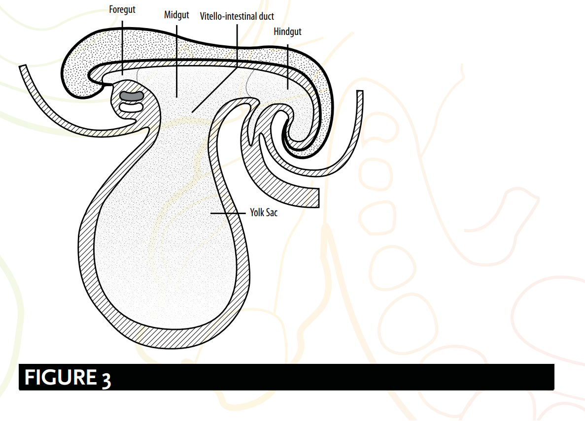
Het vouwen van de embryo is ook verantwoordelijk voor het vormen van een bloedzuigerachtige vorm [zie Figuur 3], of zoals embryologen het hebben beschreven: een cilindrische of buisvormige structuur. Embryologen Keith Moore en T. V. N. Persaud suggereren dat:
The folding of the embryo is also responsible for forming a leech-like shape [see Figure 3], or as embryologists have described; a cylindric or tube-like structure. Embryologists Keith Moore and T. V. N. Persaud suggest that:
Een belangrijke gebeurtenis bij de vorming van de lichaamsvorm is het vouwen van de platte trilaminair embryonale schijf tot een enigszins cilindrische embryo. Vouwing is het gevolg van de snelle groei van de embryo, in het bijzonder de hersenen en het ruggenmerg. 55
A significant event in the establishment of body form is the folding of the flat trilaminar embryonic disc into a somewhat cylindric embryo. Folding results from the rapid growth of the embryo, particularly the brain and the spinal cord. 55
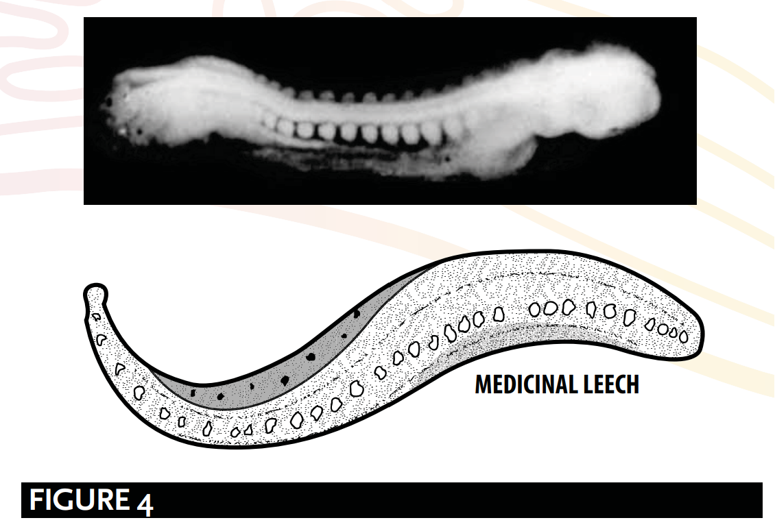
De buisvormige of bloedzuigervormige structuur [zie Figuur 4], zoals uitgelegd door Barry Mitchell en Ram Sharma, is te wijten aan:
The tube-like or leech-like structure [see Figure 4], as explained by Barry Mitchell and Ram Sharma, is due to:
longitudinale vouwing, die plaatsvindt tussen dag 21 en 24, resulteert in … het buigen van het embryo, zodanig dat de kop en de staart dichter bij elkaar worden gebracht... om een buisachtige structuur te vormen. 56
longitudinal folding, which occurs between days 21 and 24, result[ing] in … the embryo [bending] so that the head and tail are brought closer together...[to] form a tube-like structure. 56
SAMENVATTING
SUMMARY
De `alaqah-fase:
The `alaqah stage:
De embryo is via een verbindingssteel verbonden met de cytotrofoblast, alsof hij eraan hangt of is opgehangen.
The embryo is connected to the cytotrophoblast via a connecting stalk, as if it were hanging or suspended.
De vorming van de hersenen en het ruggenmerg, bekend als neurulatie; en tijdens de beginstadia van de vouwing van het embryo. Het is bij het hoogtepunt van deze processen dat het embryo op een bloedzuiger lijkt.
The formation of the brain and the spinal cord, known as neurulation; and during the initial stages of the folding of the embryo. It is upon the culmination of these processes that the embryo appears leech-like.
De embryo verkrijgt zijn voedingsstoffen via contact met de bloedvaten van de moeder. Dit weerspiegelt de werking van een aderwurm-achtige substantie die zijn voedingsstoffen via bloed verkrijgt.
The embryo obtains its nutrients via contact with the maternal blood vessels. This mirrors the action of a leech-like substance obtaining its nutrients via blood.
GALEN & `ALAQAH
GALEN & `ALAQAH
Critici verklaren dat het gebruik van het woord `alaqah een beschrijving is van de tweede fase van het menselijke embryo volgens Galenus. Hun vertaling van het woord `alaqah naar ‘bloedstolsel’ wekt de indruk van Galenus' beschrijving van deze fase:
Critics declare that the use of the word `alaqah is a description of Galen’s second stage of the human embryo. Their translation of the word `alaqah to ‘blood clot’ creates an impression of Galen's description of this stage:
Maar wanneer deze [de bloedklont] gevuld is met bloed, en het hart, de hersenen en de lever nog niet gevormd en onuitgesproken zijn, maar inmiddels wel een zekere stevigheid en aanzienlijke omvang hebben, dan is dit de tweede periode; de substantie van het foetus heeft de vorm van vlees en niet langer de vorm van zaad. 57
But when it [the clot] has been filled with blood, and heart, brain and liver are still unarticulated and unshaped yet have by now certain solidarity and considerable size, this is the second period; the substance of the foetus has the form of flesh and no longer the form of semen. 57
Er zijn een aantal zwakheden in de implicaties van deze critici:
There are a number of weaknesses with the implications of these critics:
1. Ten eerste vermeldt de Qur'an geen "hart, hersenen en lever...
1. Firstly, the Qur’an does not make a mention of the “heart, brain and liver...
“onuitgesproken en vormloos, maar toch met een zekere solidariteit en van een aanzienlijke omvang”.
[being] unarticulated and unshaped yet [with] a certain solidarity and [of a] considerable size”.
2. Ten tweede betekent het woord `alaqah niet bloed; de primaire betekenissen zijn eerder ‘een aanhangende vorm’ en ‘een aderleeider-achtige substantie’. Sommige Arabische taalkundigen zouden de betekenis van een ‘bloedstolsel’ toeschrijven aan het woord `alaqah, niet omdat het gevuld is met bloed, maar juist vanwege het feit dat het van nature ‘aanhangt’. Deze interpretatie komt overeen met de primaire betekenis van het woord.
2. Secondly, the word `alaqah does not mean blood; rather, its primary meanings are ‘a clinging form’ and ‘a leech-like substance’. Some Arab linguists would attribute the meaning of a ‘blood clot’ to the word `alaqah, not because of it being filled with blood, but instead due to it ‘clinging’ by nature. This interpretation matches the primary meaning of the word.
3. Tende, Galenus houdt vol dat "het foetus de vorm van vlees heeft" tijdens de tweede fase, terwijl de Qur'an bevestigt dat de mudghah-fase (een gekauwde stuk vlees of een klont vlees) volgt op de `alaqah-fase.
3. Thirdly, Galen insists “the foetus has the form of flesh” during the second stage, whereas the Qur’an affirms the mudghah (a chewed piece of meat or lump of flesh) stage comes after the `alaqah stage.
4. Ten tweede, in zijn boek On the Natural Faculties schrijft Galen: zo is het met het sperma: de eigenschappen die het bezat, bezat het vanaf het begin.58 Galens onjuiste implicatie dat het sperma de enige verantwoordelijke is voor het genetisch materiaal is onverenigbaar met, en staat in directe tegenstelling tot, de Qur'an en de Prophetic tradities. Zoals eerder onderzocht (zie Aristotle, Galen & Nutfah), beschouwen de islamitische bronteksten de nutfah als een gemengde druppel of vloeistof van zowel de man als de vrouw, en bevestigen zij dat zowel de man als de vrouw verantwoordelijk zijn voor de genetica van het kind.59 Daarom, als de Profeet ﷺ de visies van Galen over de menselijke ontwikkeling had gekopieerd, hoe is het dan dat hij de onjuiste wetenschap verwierp en alleen dat gebruikte wat correct was, ook al was er in het 7e-eeuwse Arabië geen enkele manier om te weten welke visies accuraat waren?
HET VERKEERD VOORSTELLEN VAN HET UITERLIJK VAN DE EMBRYO
MISREPRESENTING THE EMBRYO’S APPEARANCE
Hedendaagse commentatoren beweren dat de embryo er alleen als een bloedzuiger uitziet wanneer de dooierzak wordt verwijderd, en dat de beschrijving van de 'alaqah daarom een verkeerde weergave is van het uiterlijk van de embryo in deze fase. Embryologen leggen echter uit dat de dooierzak van de embryo wordt gescheiden door een extra-embryonale structuur, de extra-embryonale coelom genoemd. Dit feit weerlegt de claims van een verkeerde weergave, aangezien de dooierzak geen deel uitmaakt van de structuur van de embryo.60
Contemporary commentators argue the embryo only looks leech-like when the yolk sac is removed, and that the `alaqah's description is therefore a misrepresentation of the embryo’s appearance at this stage. However, embryologists explain that the yolk sac is separated from the embryo by an extra-embryonic feature, called the extra-embryonic coelom. This fact disproves claims of misrepresentation as the yolk sac is not part of the structure of the embryo.60